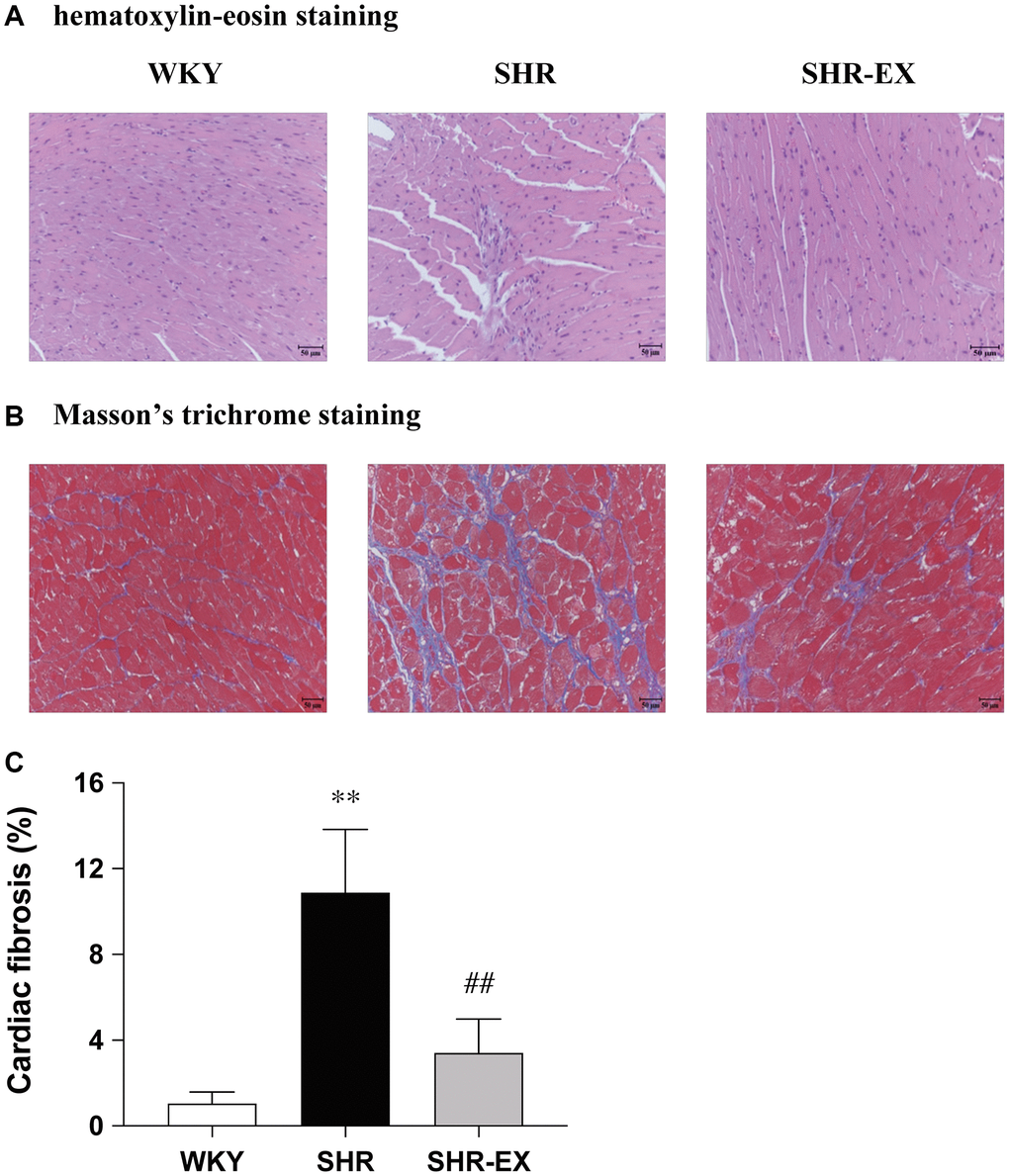
Figure 1.Representative histopathological analysis of cardiac tissue sections was performed with (A) hematoxylin-eosin staining (interstitial space: wide, arrows indicated) and (B) Masson’s trichrome staining (fibrosis: blue color, arrows indicated). The images of the myocardial architecture are magnified 200×; Bar scales = 50 μm. (C) The bar represents the percentage of blue area to the field area in Masson’s trichrome staining. Data are expressed as the mean values ± SD (n = 6 in each group). **P < 0.01 vs. the WKY group. ##P < 0.01 the SHR group vs. SHR-EX group.