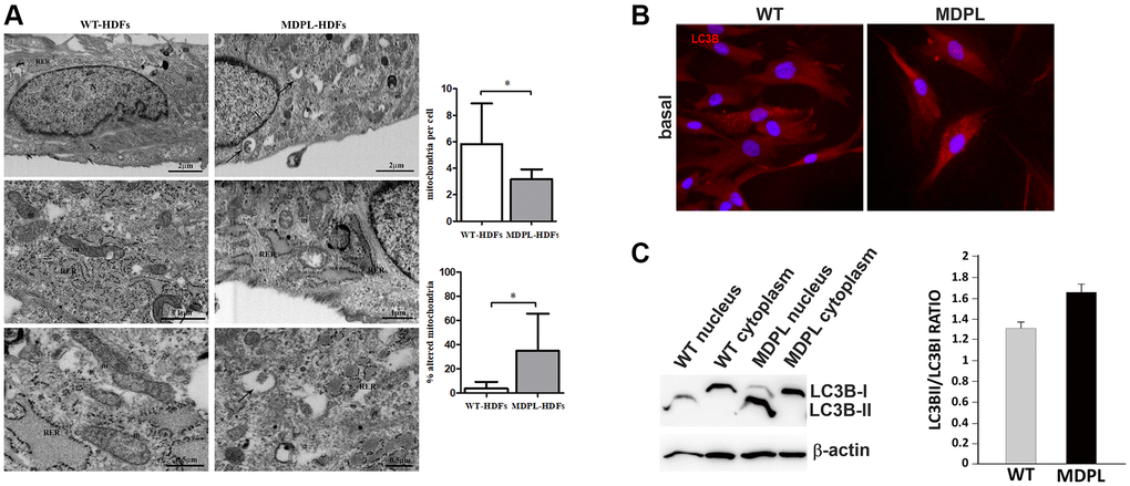
Figure 2.Ultrastructural analyses of mitochondria and their autophagic activity. (A) FIB/SEM analysis of MDPL-HDFs vs. WT fibroblasts. Healthy cells (on the left) show regular nucleus (N), several mitochondria (m) and abundant rough endoplasmic reticulum (RER). By contrast, MDPL-HDFs cells (on the right) display fewer RER cisternae, while smooth endoplasmic reticulum (SER) and Golgi apparatus are more prominent than in WT. Diseased cells also show several autophagosomes (black arrows), often containing partially digested mitochondria. Statistical analysis demonstrates significant decreased number of mitochondria, which are also significantly more damaged, than their normal counterpart (*p < 0.05, for both parameters). (B) Representative image of immunofluorescence analysis of LC3 in WT and MDPL HDFs. (C) Western blot densitometric analysis of the LC3-II/I ratio. Data are presented as means ± SD. β-actin was used as control.