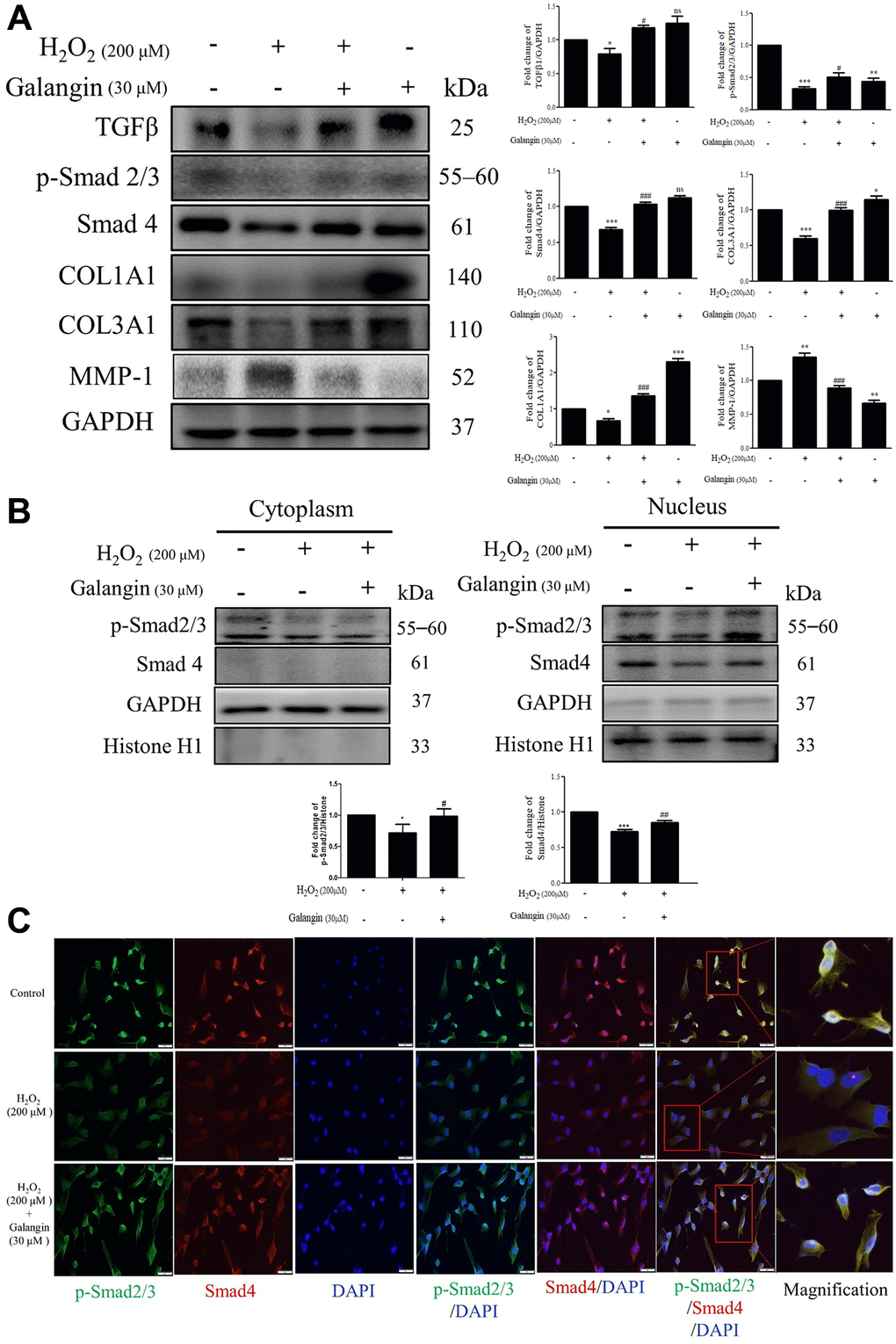
Figure 4.Galangin attenuates H2O2-induced TGFβ/Smad collagen synthesis pathway impairment in HS68 cells. (A) HS68 cells were exposed to H2O2 (200 μM) for 1 h and then treated with galangin (30 μM) for 23 h. Protein expression of collagen synthesis-related pathway components (TGFβ, p-smad2/3, Smad4, COL1A1, COL3A1) and collagen degradation-related protein (MMP-1) were detected by western blot. (B) The protein expression of p-smad2/3, Smad4 in the nuclear and cytosolic fractions was detected by western blotting. GAPDH was used as a loading control. Values are shown as mean ± SE. Quantification of the results is shown (n = 3) *P < 0.05, **P < 0.01, ***P < 0.001 vs. untreated control cells; #P < 0.05, ##P < 0.01, ###P < 0.001 vs. H2O2-treated cells. (C) Anti-p-Smad2/3, Smad4 antibody, and FITC/PE-conjugated secondary antibody were used to detect p-Smad2/3 (green), Smad4 (red) expression. DAPI indicated the nucleus location (blue). The images were captured using a florescence microscope (200×).