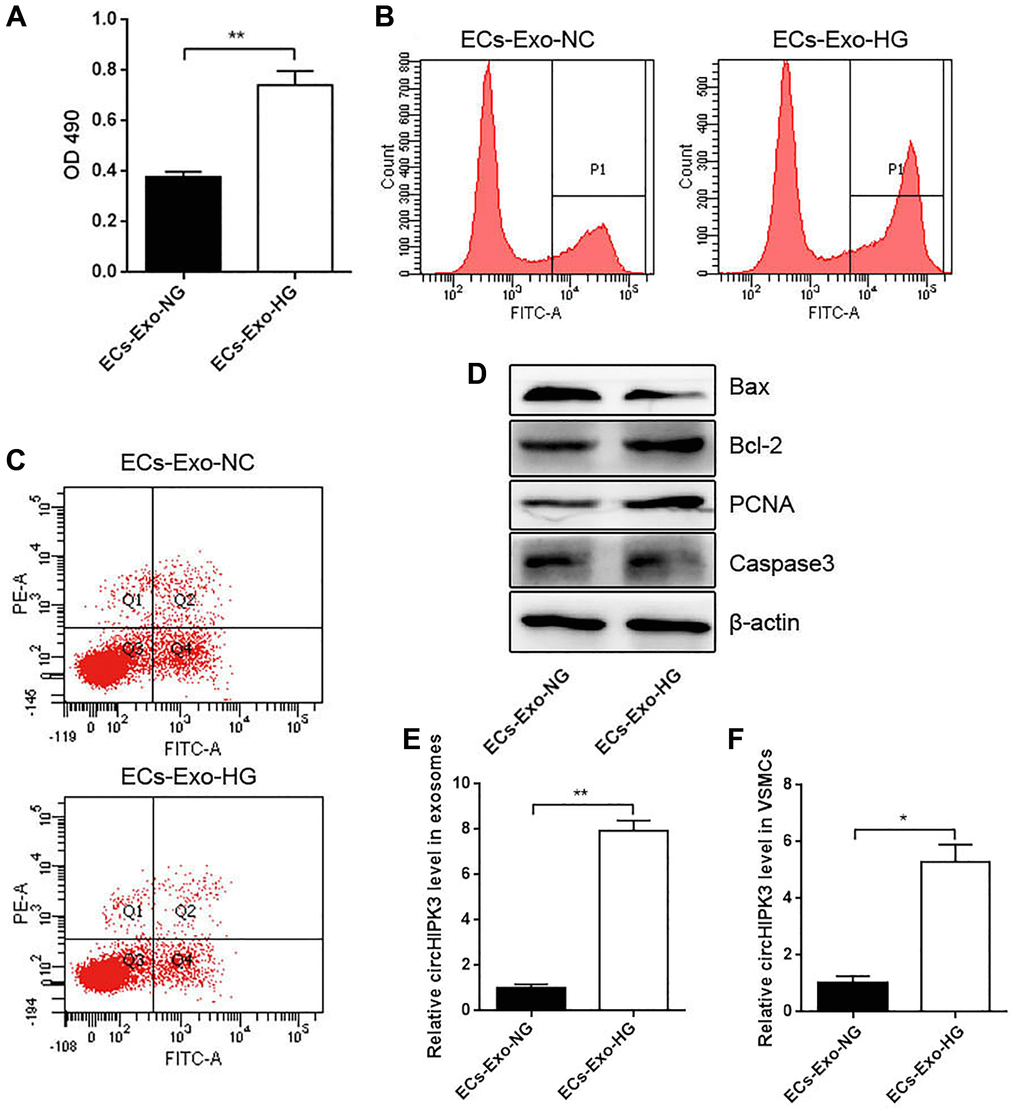
Figure 2.Effects of MAEC-derived exosomes on VSMC proliferation and apoptosis. (A) CCK-8 was used to detect cell viability in VSMCs incubated with exosomes isolated from MAECs cultured in an HG or NG medium (**p < 0.01 ECs-Exo-NG vs. ECs-Exo-HG). (B) Edu assay was used to detect cell viability by FCM. (C) FCM detected the apoptosis of VSMCs. (D) Western blot analysis detected the expression of Bax, Bcl2, PCNA, and Caspase 3. (E) The relative expression of circHIPK3 in the exosomes of ECs-Exo-NG or ECs-Exo-HG detected by qRT-PCR (**p < 0.01 ECs-Exo-NG vs. ECs-Exo-HG). (F) The relative expression of circHIPK3 in VSMCs incubated with ECs-Exo-NG or ECs-Exo-HG detected by qRT-PCR (*p < 0.05 ECs-Exo-NG vs. ECs-Exo-HG).