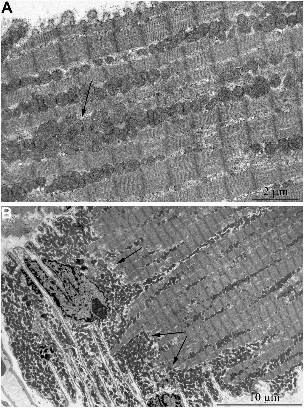
Figure 4.Ultrastructure of mitochondria in skeletal muscle of 5-year-old naked mole rat. (A) Longitudinal section of muscle fiber. Rows of mitochondria arranged along myofibrils can be observed, mitochondrial cluster is indicated by arrow. (B) Longitudinal section of muscle fiber. Large clusters of mitochondria in the perinuclear and subsarcolemmal areas are indicated by arrows.