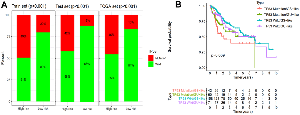
Figure 6.Comparison of the GILncSig with TP53 mutation status in prognosis value. (A) The proportion of TP53 mutation in high- and low-risk groups in the training set, testing set and the TCGA set. (B) Kaplan–Meier curve analysis of overall survival is shown for patients classified according to TP53 mutation status and the GILncSig. Statistical analysis was performed using the log-rank test.