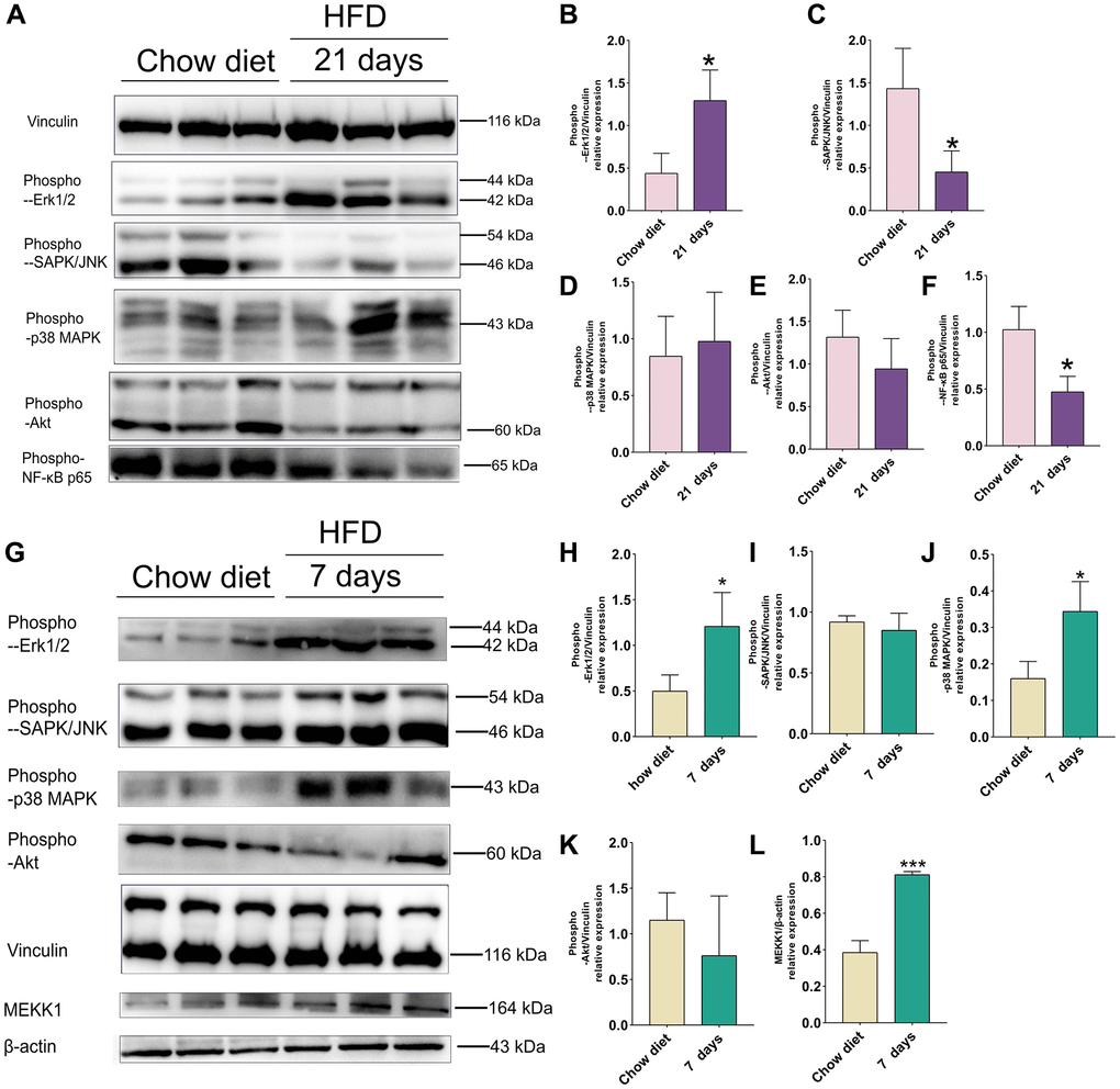
Figure 5.Effect of HFD on signaling pathways in the mice cortex at 21 days and cerebellum at 7 days. (A) Western blots and (B–F) Quantitation for phospho-p44/42 MAPK (Erk1/2) (B), phospho-SAPK/JNK (C), phospho-p38 MAPK(D), phospho-Akt (E), phospho-NF-κB p65 (F) in the cortex of mice after 21 days HFD (n = 3 per group). (G) Western blots and (H–L) Quantitation for phospho-p44/42 MAPK (Erk1/2) (H), phospho-SAPK/JNK (I), phospho-p38 MAPK (J), phospho-Akt (K), MEKK1 (L) in the cerebellum of mice after 7 days HFD (n = 3 per group). Vinculin and β-actin as a loading control. Values are presented as means ± SD. *P < 0.05 and **P < 0.01 versus chow diet; Two-tailed Student’s t test.