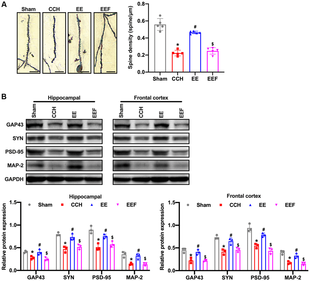
Figure 4.EE improves synaptic morphology in CCH rats. (A) Neuronal spine density changes were detected by Golgi staining in the hippocampal in each group. Western blot were used to detect the expression of synaptic plasticity associated proteins. Number and length of dendritic spines. N = 5. (B) Levels of GAP43, SYN, PSD-95, MAP-2. *p < 0.05, vs. Sham group; #p < 0.05, vs. CCH group. $p < 0.05, vs. EE group. N = 3. Sham group, treated with an equal volume of vehicle; CCH group, chronic cerebral hypoperfusion and no treatment; EE group, CCH and treated with EE. EEF group, CCH and treated with EE and FSH. *p < 0.05, #p < 0.05, $p < 0.05.