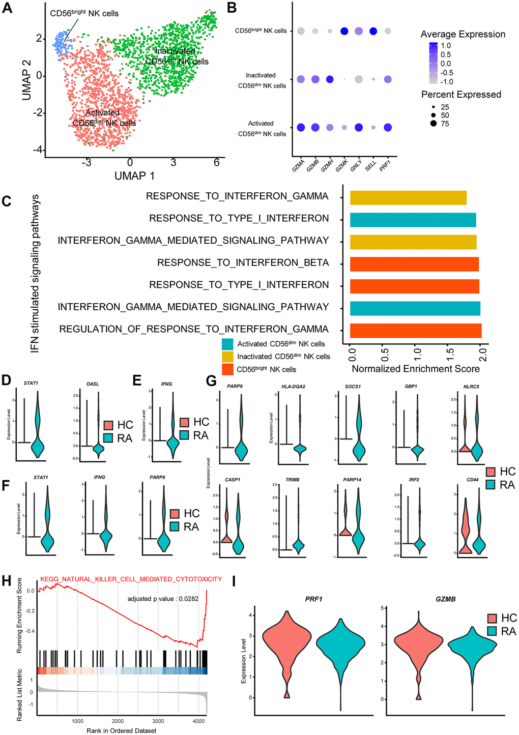
Figure 3.Interferon (IFN)-stimulated pathways promote cytokine secretion and inhibit cytotoxicity in rheumatoid arthritis (RA) natural killer (NK) cells. (A) Two-dimensional uniform manifold approximation and projection (UMAP) visualization of reclustered NK cells. Three NK cell clusters (activated CD56dim NK cells, inactivated CD56dim NK cells and CD56bright NK cells) were identified. (B) Dot plot illustrating the expression levels of several marker genes in three NK cell subtypes. (C) Bar plots of selected gene set enrichment analysis (GSEA) results indicated altered IFN signaling pathways in three NK cell subtypes. (D) Violin plots of significantly upregulated type I IFN-stimulated genes in RA-activated CD56dim NK cells (STAT1) and in RA CD56bright NK cells (OASL). (E) Violin plot of significantly upregulated IFN-γ-stimulated genes in RA CD56bright NK cells. (F) Violin plots of significantly upregulated IFN-γ-stimulated genes in RA-activated CD56dim NK cells. (G) Violin plots of significantly upregulated IFN-γ-stimulated genes in RA-inactivated CD56dim NK cells. (H) GSEA plot of the “KEGG_NATURAL_KILLER_CELL_MEDIATED_CYTOTOXICITY” pathway in RA inactivated CD56dim NK cells. (I) Violin plots of significantly upregulated cytotoxic effector genes in RA activated CD56dim NK cells. All upregulated genes satisfied log2 (fold change)>0.25 and adjusted p-value<0.05.