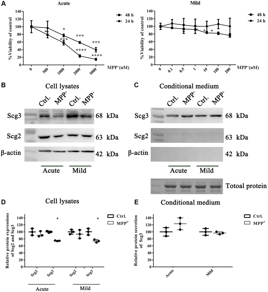
Figure 3.The effects of acute and mild MPP+ treatments on the cell viability and expressions of secretogranins in SH-Sy5Y cells. (A) SH-Sy5Y cells were exposed to an acute (500–5000 μM) and a mild (0.1–200 μM) treatment of MPP+ for 24 and 48 h. The cell viability was measured using the MTT assay. And appropriate treatments were determined as 1000 μM for 24 h and 10 μM for 48 h, respectively. Data from three individual experiments were expressed as the mean percentage of the controls. (B) Intracellular protein expressions of Scg2 and Scg3 in the dopaminergic SH-sy5Y cells under acute and mild MPP+ treatments were analyzed by immunoblotting and quantified by densitometric analysis normalized to β-actin. (C) The secretions of Scg2 and Scg3 in the conditioned medium of SH-sy5Y cells were analyzed by immunoblotting. According to the Coomassie staining, an equal amount of total protein in the conditioned medium was loaded for each sample. (D) The quantifications of intracellular Scg2 and Scg3 levels in the dopaminergic SH-sy5Y cells under acute and mild MPP+ treatments. (E) The quantifications of Scg3 secretion levels in the dopaminergic SH-sy5Y cells under acute and mild MPP+ treatments. Two-tailed unpaired Student t-tests were performed between the control and treated groups. *Statistically significant with P < 0.05; Error bars are SD; N = 3.