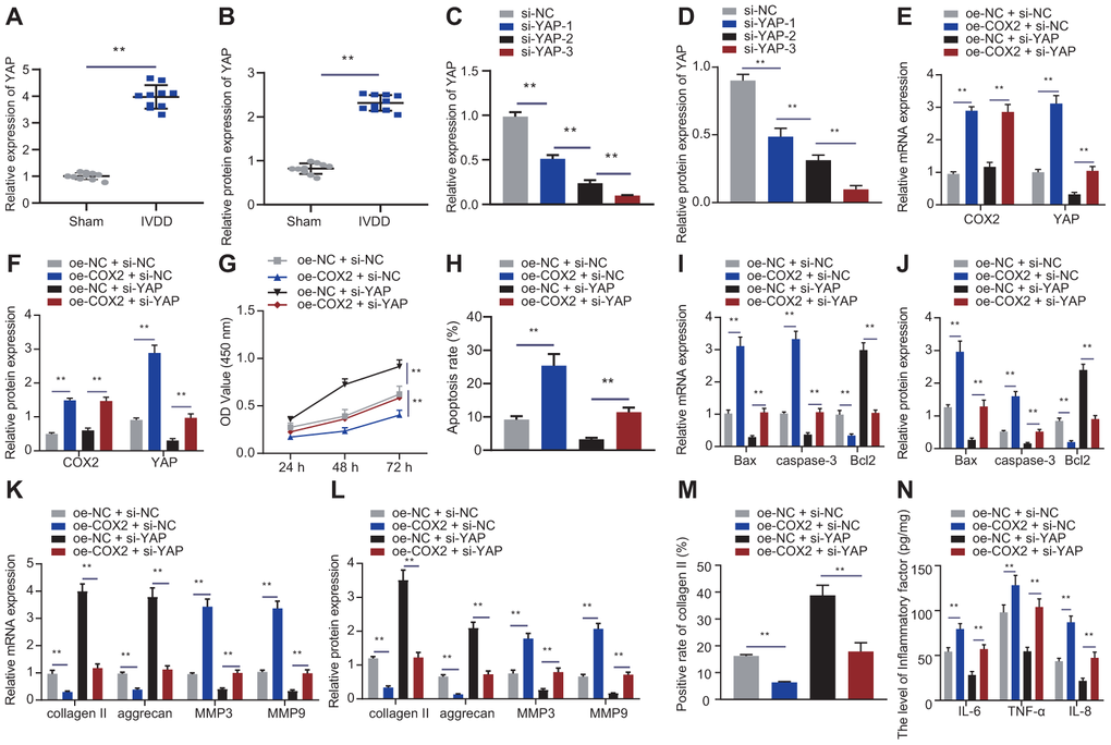
Figure 6.COX2 promoted YAP/Hippo signaling pathway to promote NP cell proliferation. (A) The expression of YAP in the intervertebral disc tissue of IVDD and sham-operated rats was detected by RT-qPCR (n = 10). (B) Western blot showing the expression of YAP in the intervertebral disc tissue of IVDD and sham-operated rats. (C) The interference efficiency of si-YAP in NP cells after 24 hours of transfection was detected by RT-qPCR. (D) The interference efficiency of si-YAP in NP cells after 48 hours of transfection was detected by Western blot. (E) mRNA expression of COX2 and YAP in NP cells after 24 hours of transfection with oe-COX2 and si-YAP was detected by RT-qPCR. (F) COX2 and YAP protein expression in NP cells after 48 hours of transfection with oe-COX2 and si-YAP was detected by Western blot. (G) The proliferation of NP cells after 24 hours of transfection with oe-COX2 or si-YAP was detected by CCK-8. (H) The apoptosis of NP cells after 48 hours of transfection with oe-COX2 or si-YAP was detected by flow cytometry. (I) The expression of apoptosis-related factors Bax, Bcl-2, and caspase-3 was detected by RT-qPCR in NP cells after 24 hours of transfection with oe-COX2 or si-YAP. (J) The expression of apoptosis-related factors Bax, Bcl-2, and caspase-3 was detected by Western blot in NP cells after 48 hours of transfection with oe-COX2 or si-YAP. (K) The expression of collagen II, aggrecan, MMP3, and MMP9 was detected by qRT-PCR in NP cells after 24 hours of transfection with oe-COX2 or si-YAP. (L) The expression of collagen II, aggrecan, MMP3, and MMP9 was detected by Western blot in NP cells after 48 hours of transfection with oe-COX2 or si-YAP. (M) Immunofluorescence staining showing collagen II protein in NP cells after 48 hours of transfection with oe-COX2 or si-YAP. (N) Inflammatory factors IL-6, TNF-α, IL-8 levels in NP cells after 48 hours of transfection with oe-COX2 or si-YAP were detected by ELISA. Measurement data are expressed as the mean ± standard deviation (n = 3) and analyzed using independent sample t-tests between two groups, using one-way ANOVA between multiple groups, or using two-way ANOVA between groups at different time points. **, p < 0.01.