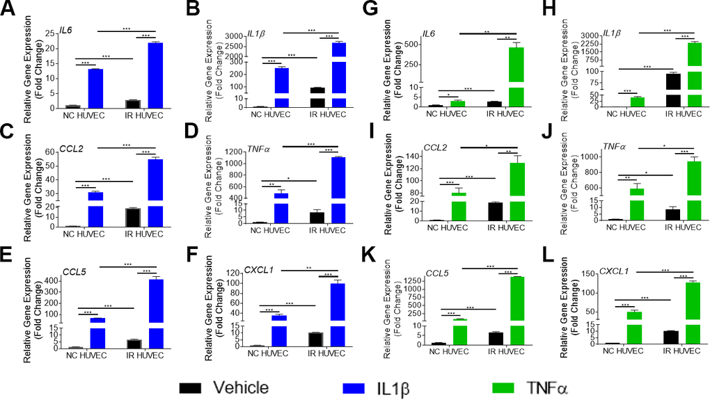
Figure 2.Comparison of the SASP gene expression in IL1β and TNFα-stimulated NC HUVEC and IR HUVEC. Relative fold change in gene expression of IL6 (A), IL1β (B), CCL2 (C), TNFα (D), CCL5 (E), and CXCL1 (F) in NC HUVEC and IR HUVEC 3 hours after stimulation with 3 ng/mL IL1β. Relative fold change in gene expression of IL6 (G), IL1β (H), CCL2 (I), TNFα (J), CCL5 (K), and CXCL1 (L) in NC HUVEC and IR HUVEC 3 hours after stimulation with 3 ng/mL TNFα. Gene expression in unstimulated NC HUVEC was used as baseline and GAPDH was used as endogenous control. (n = 3; mean ± SEM; * p<0.05, ** p<0.01, *** p<0.001).