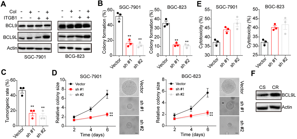
Figure 3.BCL9L was up-regulated in 3D collagen cultured ITGB1+ gastric cancer. (A) The ITGB1-/+ SGC-7901 cells were sorted and cultured in 3D collagen gel (6 days) or not. The expression of BCL9, BCL9L and β-actin was examined by western blotting. (B) The ITGB1+ SGC-7901 cells were sorted and cultured in 3D collagen gel (6 days). Then tumor cells were treated with BCL9L shRNA or vector and the 3D colony formation capability was examined. (C) The ITGB1+ SGC-7901 cells were sorted and cultured in 3D collagen gel (6 days). Then tumor cells were treated with BCL9L shRNA or vector and the tumorigenic capability was examined. (D) The colony sizes of tumor cells in (C). The scale bar is 30 μm. (E) The ITGB1+ SGC-7901 cells were sorted and cultured in 3D collagen gel (6 days). Then tumor cells were treated with BCL9L shRNA or vector. Then tumor cells were treated with 5-FU (5 μg/ml) and the cell apoptosis was examined. (F) Western blotting of BCL9L and β-actin in chemo-sensitive (CS) and chemo-resistant (CR) tissues from gastric patients. *Indicates P <0.05, ** Indicates P <0.01.