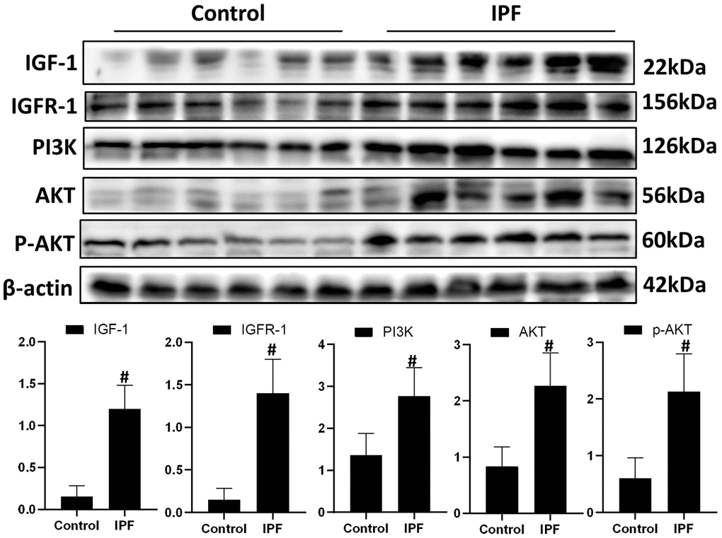
Figure 1.Activated IGF1 pathway in lung tissue from patients with IPF. IGF-1, IGFR-1, PI3K, AKT and p-AKT were assessed using western blotting analyses. #P < 0.01 for the comparison between the control group with the IPF group. Unpaired, two-tailed Student’s t test.