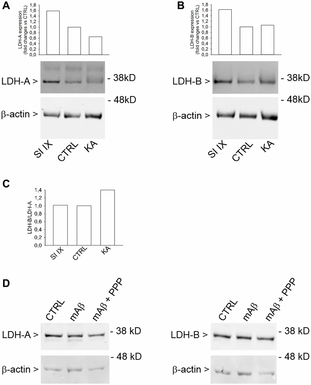
Figure 8.Increased LDH-B/LDH-A expression ratio in neurons challenged with kainate. Western blot analysis of LDH-A (A) and LDH-B (B) in lysates obtained from neurons that were deprived from glucose for 2 hr before returning to 3 mM glucose. Kainate (KA, 100 μM for 40 min) reduced LDH-A (A) without affecting LDH-B expression (B). γ-Secretase inhibitor IX (SI IX), 100 nM during glucose deprivation and for 40 min following glucose re-addition, increased both LDH-A (A) and LDH-B (B) expression. In (A and B), graph bars represent fold changes of LDH-A and LDH-B over the respective control (CTRL). Densitometry signals were normalized on β-actin. In (C), graph bars represent the ratio between LDH-B and LDH-A values as expressed in (B and A), respectively. The experiment was repeated twice with similar results. Hybridization signals were detected with the Odyssey infrared imaging system in their original green or red colors and automatically converted into greyscale. In (D), western blot images of LDH-A and LDH-B in lysates from neurons that, following glucose deprivation and replenishing, were exposed to Aβ42 monomers in the absence (mAβ, 100 nM for 40 min) and in the presence of 500 nM PPP (mAβ + PPP). None of the treatments modified LDH-A or LDH-B expression.