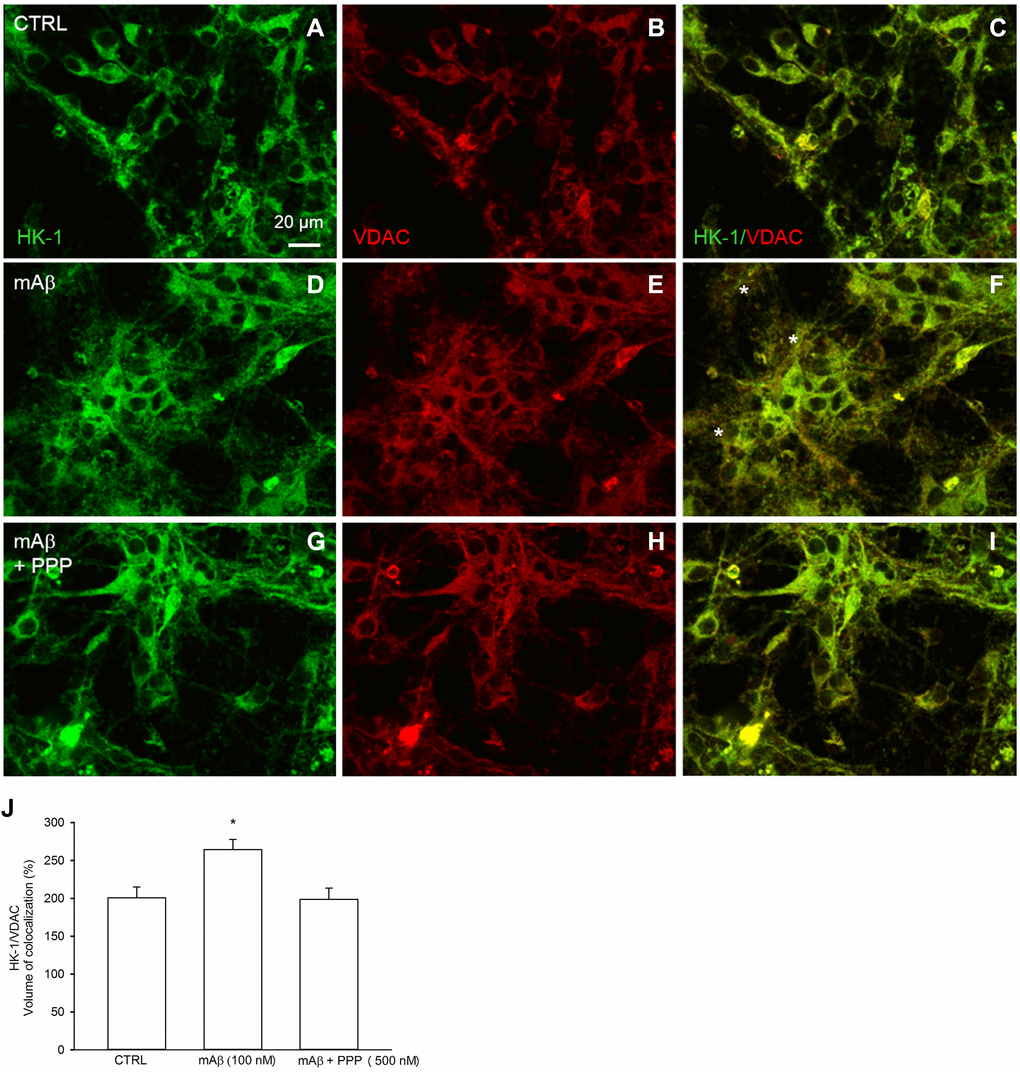
Figure 5.Synthetic Aβ42 monomers increased the mitochondrial localization of HK-1 at the neurite processes in a manner dependent on IGF-IR activation. Confocal images of primary cortical neurons glucose-starved for 2 hours before returning to 3 mM glucose, in the absence (CTRL, A–C) or in the presence of either 100 nM synthetic Aβ42 monomers for 40 min (mAβ, D–F) or synthetic Aβ42 monomers + 500 nM PPP (mAβ + PPP, G–I). Neurons were immunostained for HK-1 (green fluorescence) and VDAC (red fluorescence). Overlays of green and red fluorescence for each experimental conditions are shown on the right side of the figure (C, F, I). In (F) asterisks indicate neurite processes exhibiting green (HK-1)/red (VDAC) co-localization (orange to yellow). Images were not altered in any way, but were despeckled by ImageJ to reduce noise. Scale bar = 20 μm. In (J), bars represent the % image volume colocalized (i.e., the percentage of voxels which have both green (HK-1) and red (VDAC) fluorescence intensity above the threshold with respect to the total number of pixels in the image) for each experimental conditions, and values are expressed as means ± S.E.M. of 3 determinations. Each determination represented a culture dish in which the % of image volume colocalized was calculated from three random fields. *p < 0.001 vs. control (CTRL); one-way ANOVA with post hoc Holm-Sidak multiple comparisons vs. control group.