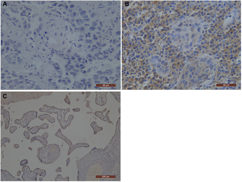
Figure 1.Representative photographs of PD-L1 immunostaining in esophageal squamous cell carcinoma. The positive staining was assessed against the positive control staining (Placental tissue). (A) Negative immunohistochemical staining pattern for PD-L1; (B) Positive immunohistochemical staining pattern for PD-L1; (C) the positive control staining (Placental tissue); PD-L1, programmed death-ligand 1.