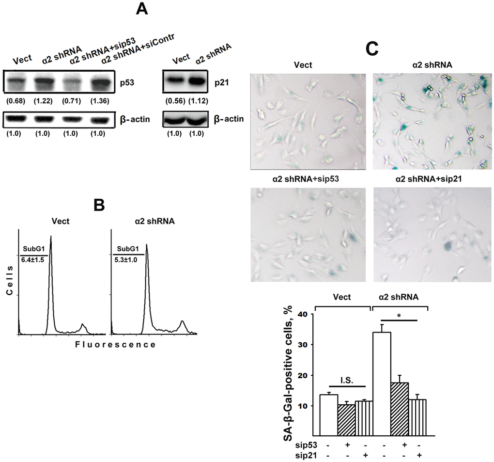
Figure 2.Silencing of p53 and p21 reversed the stimulatory effect of α2β1 knockdown on senescence of SK-Mel-147 melanoma cells. The cells were transduced with the appropriate vectors and transfected with p53- or p21-specific siRNAs as described in Materials and Methods. (A) Western-blotting of the cellular lysate proteins. The procedures were performed as described in Materials and Methods and the legend to Figure 1. Numbers below the bands indicate the p53 or p21 band densities normalized against β-actin. Shown are representative blots. (B) Apoptosis assay. Cells transduced with the appropriate vectors were stained with propidium iodide, analyzed by flow cytometry (Materials and Methods) and the percentage of cells with subG1 DNA was determined. Shown are representative histograms; SubG1 values were derived from three independent experiments (M ± SEM). (C) SA-β-Gal staining. The cells were treated and counted as described in Materials and Methods and in the legend to Figure 1; magnification: × 200. The results of three independent experiments are shown (M ± SEM); *, ρ < 0.05. Vect, scramble shRNA transduced cells; α2 shRNA, α2 shRNA transduced cells; I.S, insignificant.
Figure 2 — Implication of integrin α2β1 in senescence of SK-Mel-147 human melanoma cells | Aging