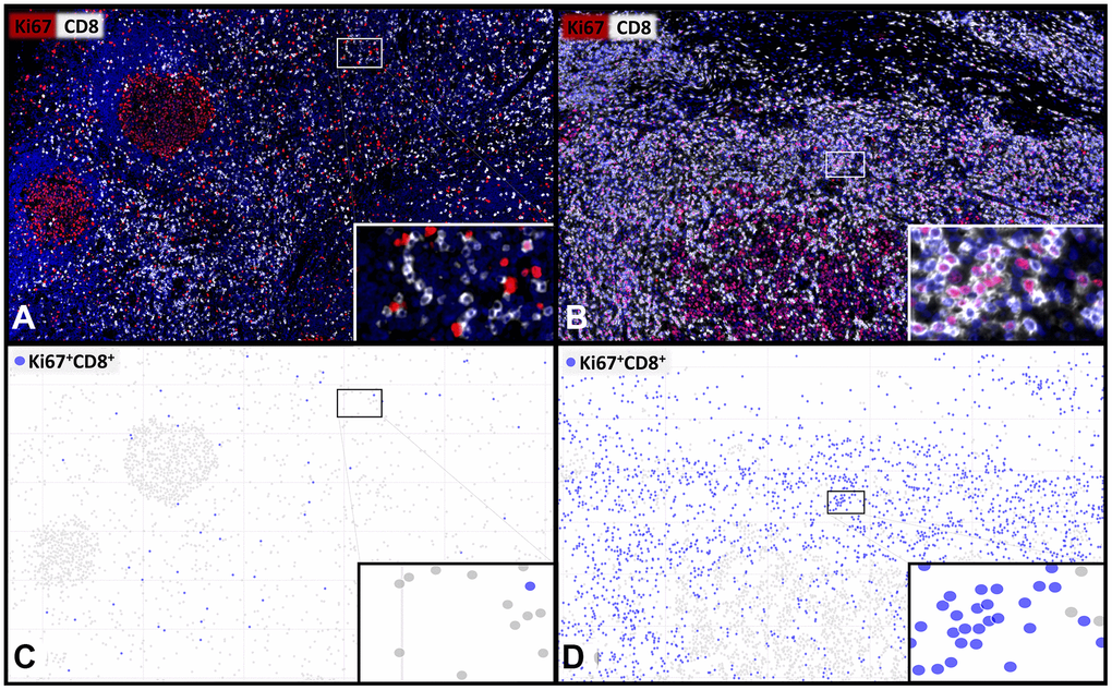
Figure 1.Representative images and visualisations showing the cell detection of proliferating CD8+ T-lymphocytes. Multiplex immunofluorescence images (A, B) showing CD8+ cytotoxic T-lymphocytes (white) and Ki67+ proliferating cells (red) with a low (A, C) and a high (B, D) percentage of proliferating Ki67+CD8+ T-cells. The visualization (C, D) of the digital image analysis underlines the accuracy of the automated detection of the subset of Ki67+CD8+ proliferating cytotoxic T-cells (blue). 400x magnifications are shown in the insets.