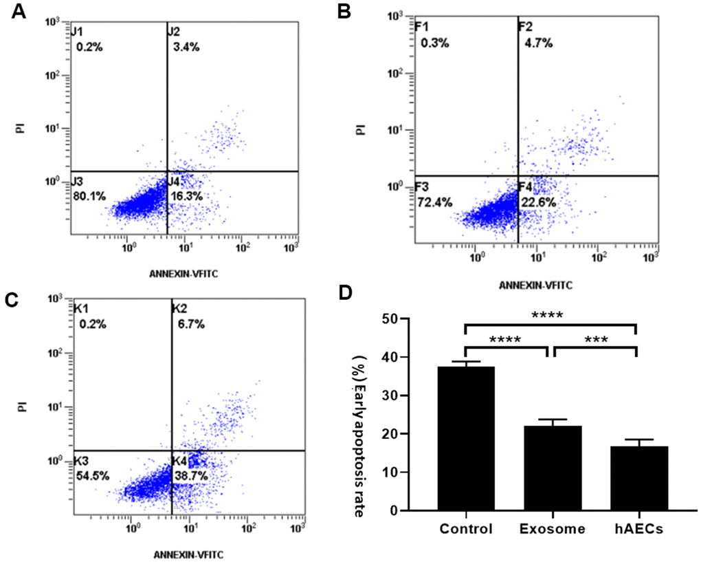
Figure 7.The apoptosis rate of cardiomyocytes was measured by flow cytometry. The results showed that both hAECs and exosomes decrease apoptosis. (A) Treatment with hAECs. (B) Treatment with exosomes. (C) Control group. (D) The bar graph shows quantitative analysis of early apoptosis. The data are shown as the means ± SD, n=4, *P<0.05.