Submit an Article
Navigate
Home
Editorial Board
Editorial Policies
Current Volume
Archive
Scientific Integrity
Publication Ethics Statements
Interviews with Outstanding Authors
Newsroom
Sponsored Conferences
Podcast
Contact
Special Collections
Submit an Article
Online ISSN: 1945-4589
Research Paper
|
Volume 13, Issue 12
|
pp. 15964–15989
Antrodia salmonea
induces apoptosis and enhances cytoprotective autophagy in colon cancer cells
Back to article
Figure 14
(14 of 15)
−
100%
+
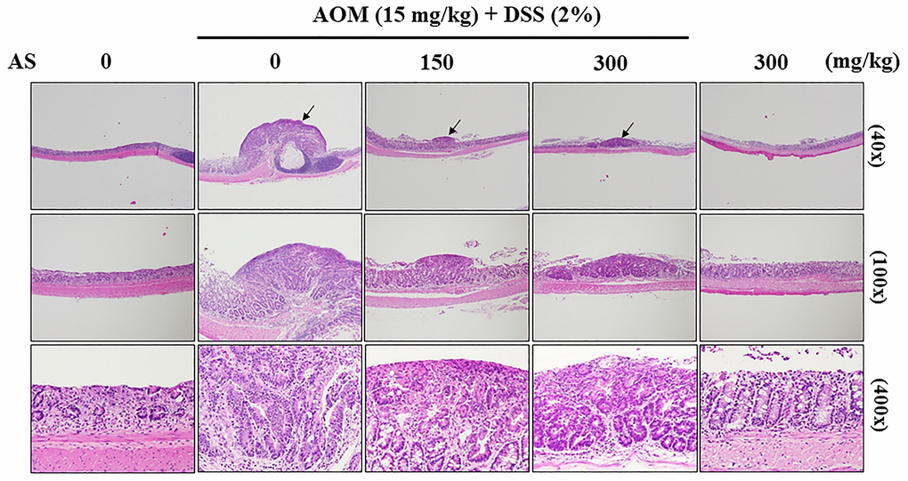
Figure 14.
Histopathological examination of the colon from the AOM/DSS- and/or AS-treated ICR mice.
Representative portion of colon tissues was stained by hematoxylin and eosin, and observed under 40×, 100×, and 400× magnification.