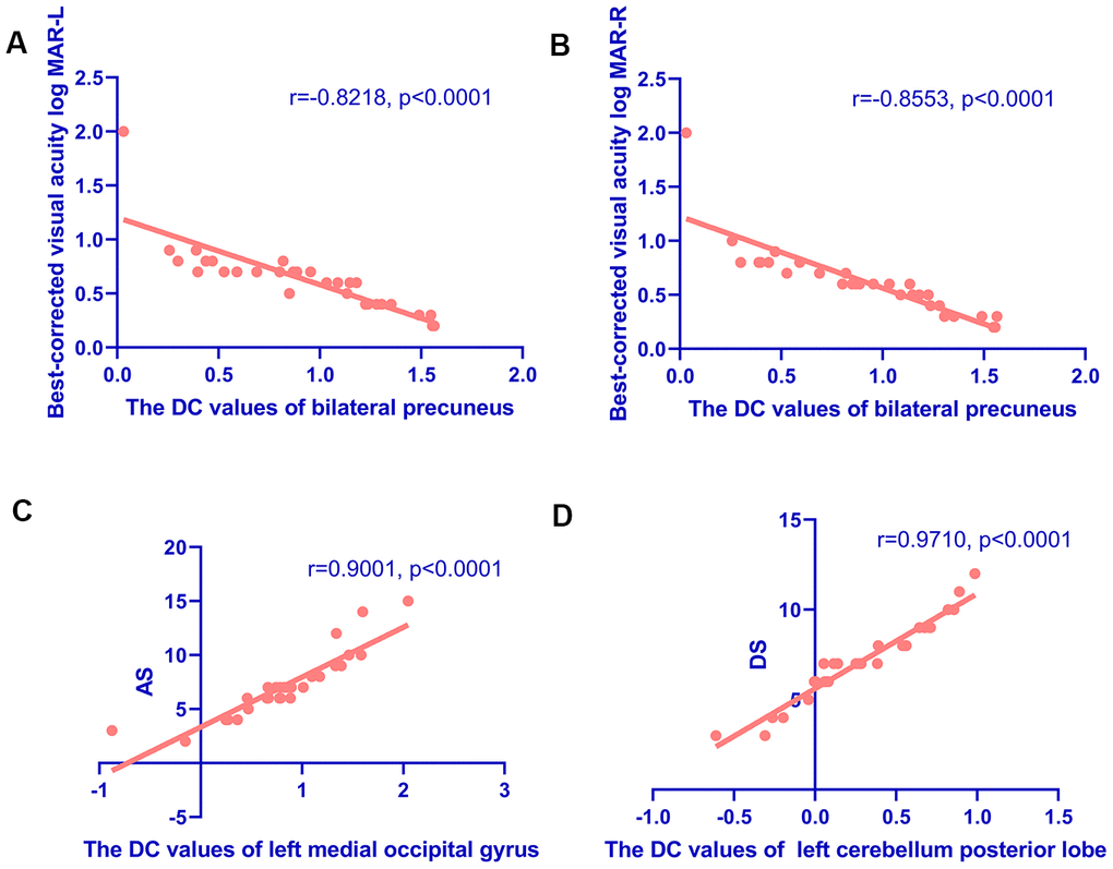
Figure 4.Correlations between the DC values of different regions and the clinical behaviors in HR group. (A, B) Correlations between the DC values of bilateral precuneus and best-corrected visual acuity. The DC values of bilateral precuneus were positively correlated with the values of BCVA-L(LogMAR) (r=-0.8218, p<0.0001) (A) and BCVA-R (LogMAR) (r=-0.8553, p<0.0001) (B). (C, D) Correlations between the DC values of specific cerebral regions and the Hospital Anxiety and Depression Scale. The DC values of the left middle occipital gyrus were positively correlated with AS (r=0.9001, p<0.0001) (C); and the DC values of the left cerebellum posterior lobe were positively correlated with the DS (r=0.9710, p<0.0001) (D). Abbreviations: DC, degree centrality; AS, anxiety scores; DS, depression scores.