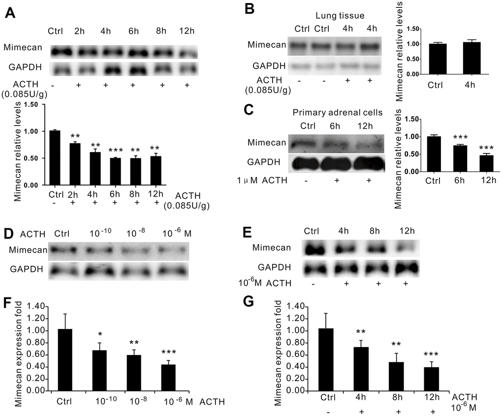
Figure 3.Inhibition of mimecan expression in time- and dose-dependent manners by ACTH administration. (A) Mimecan mRNA expression was detected by Northern blot analysis in the adrenal glands of C57BL/6 male mice after intraperitoneal injection of adrenocorticotropic hormone (ACTH; 0.085 U/g; 12 mice per group for each time point), showing the significant time-dependent decline in the ACTH injecting group compared with the control group that was not injected. (B) No change in mimecan expression was observed in the lung tissue after ACTH dosing. (C) Northern blot analysis showing that the expression of mimecan in cultured primary adrenal cells decreased at various time points after the administration of 1 μM ACTH (6 or 7 mice for each time point). (D, E) Northern blot analysis showing ACTH-induced dose-dependent (D) and time-dependent (E) inhibition of mimecan mRNA in Y-1 cells. The ACTH dose in (E) was 10-6 M. Y-1 cells were serum-deprived overnight before the addition of ACTH. (F, G) Realtime PCR showing ACTH-induced dose-dependent (F) and time-dependent (G) inhibition of mimecan mRNA in Y-1 cells. Relative mimecan mRNA levels were normalized to GAPDH mRNA expression and compared with untreated controls. Data information: *p<0.05, **p<0.01, ***p<0.001 for ACTH treatment vs. control, Student’s t-test.