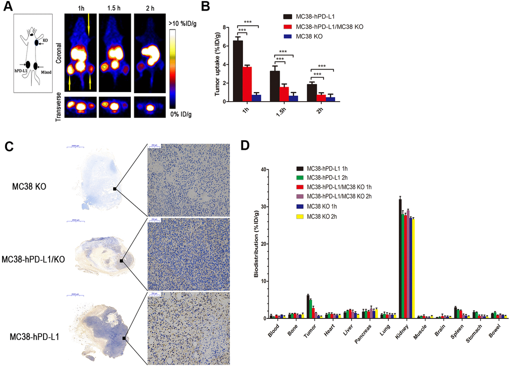
Figure 4.PET imaging of tumor-bearing models. (A) Static images at 1, 1.5, and 2h after injection of 2.5 MBq of 68Ga-NOTA-Nb109 (n=3, tumors indicated by the yellow arrow). (B) The uptake of 68Ga-NOTA-Nb109 in tumors according to quantification analysis of PET images. (C) PD-L1 immunohistochemical staining of tumors (n=5, scale bar =2 mm in the left column and 200 μm in right column). ***P <0.001. (D) Biodistribution of 68Ga-NOTA-Nb109 in major organs at 1 and 2 h (n=5).