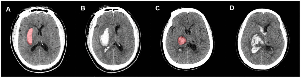
Figure 5.Representative illustration of new IVH, IVH expansion, and manual region of interest (ROI) segmentation. Images (A, B) are non-contrast CT images (axial view) of a 54-year-old male who experienced a new IVH. There was no baseline IVH (A), but the hematoma broke into ventricles on the follow-up CT (B). Images (C, D) are non-contrast CT images (axial view) of a 68-year-old female who experienced IVH expansion; (C) shows an initial IVH with a volume of 2.53 mL; follow-up CT (D) shows that the volume of IVH increased to 22.31 mL within 72 h.