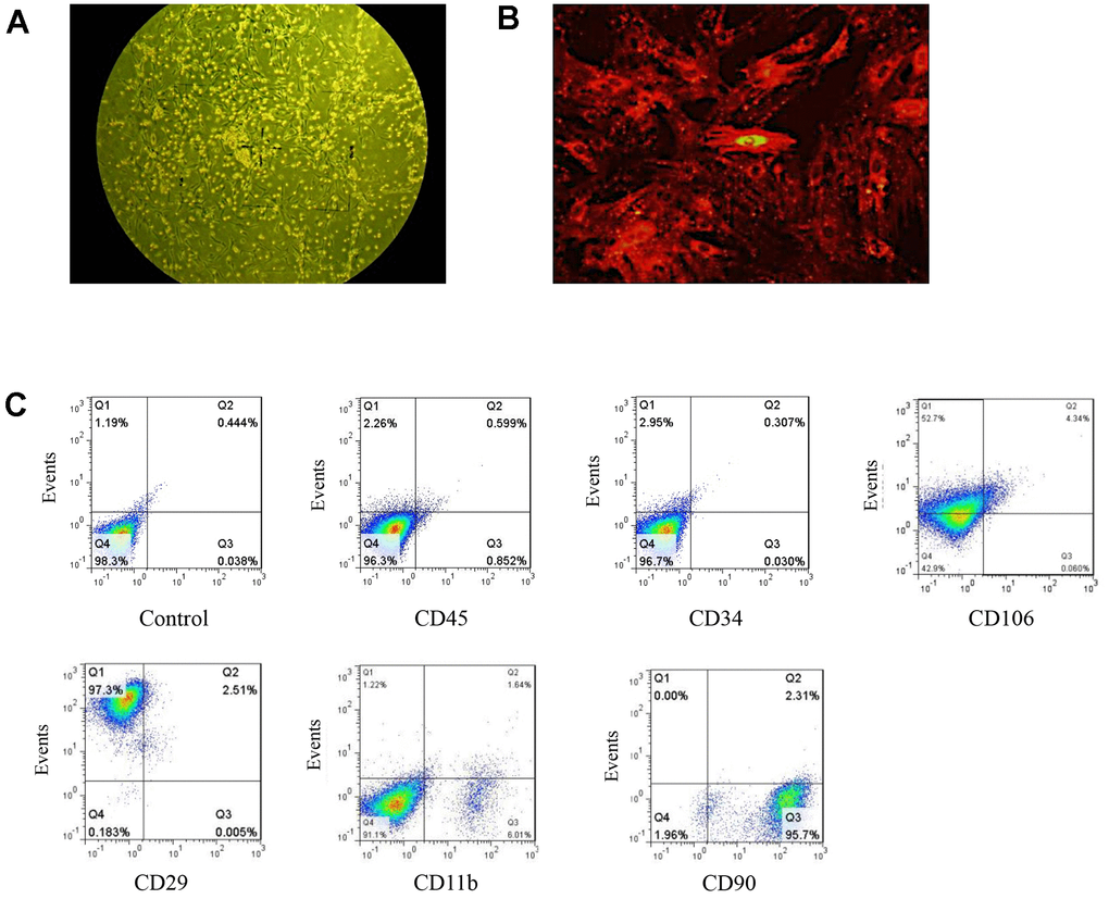
Figure 1.Isolation and identification of BMSCs. (A) BMSCs as observed under ordinary light microscopy (magnification: 100×). (B) DM-cil labeled BMSCs as observed by fluorescence microscopy (magnification: 400×). (C) The quantification of BMSC specific markers.