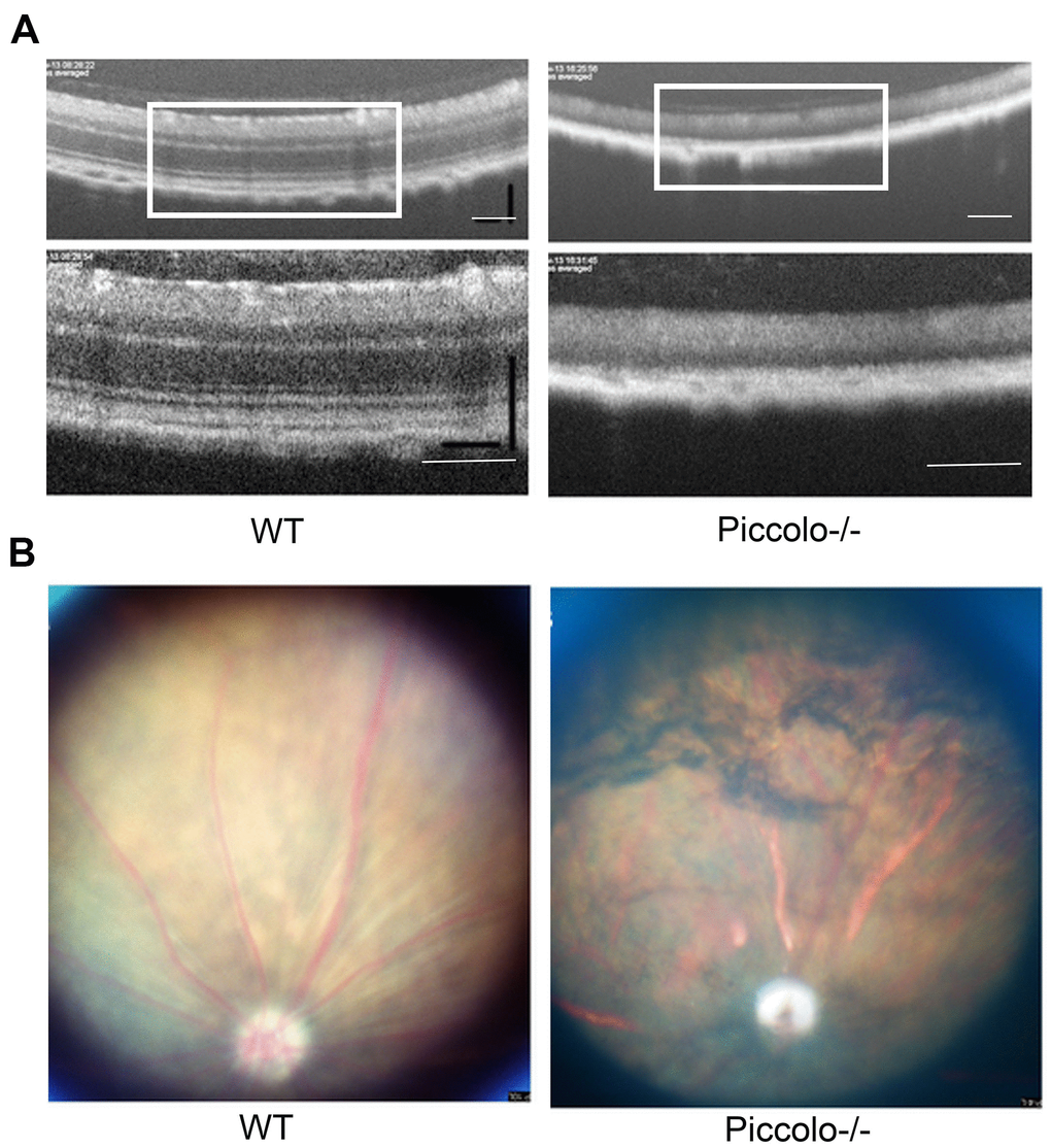
Figure 6.Examination of the retinal fundus in Piccolo-/- mice. (A) Optical coherence tomography (OCT) of the retina in Piccolo-/- mice compared with the wild type (WT) mice at P30; Scale bars: 100 μm. (B) Retinal fundus imaging in one-month-old WT and Piccolo-/- mice.