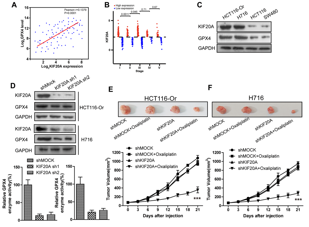
Figure 2.High KIF20A expression in resistant CRC cell line suppressed the intracellular ferroptosis process. (A) Correlation between expression levels of KIF20A and GPX4 in colorectal cancer samples. (B) The expression level of KIF20A of colorectal cancer patients in different stages. (C) The expression level of KIF20A in different colorectal cancer cell lines were examined by WB assay. (D) WB assay was used to observe whether KIF20A silencing could impact the intracellular GPX4 expression level. Top, HCT116-Or cells. Bottom, H716 cells. (E, F) HCT116-Or (E) and H716 (F) cells were selected to construct the subcutaneous xenograft model of nude mice, so as to observe whether KIF20A silencing would affect the suppression of oxaliplatin on colorectal cancer in vivo. Top, representative images of xenografted tumor in the indicated groups. Bottom, statistical results of growth of xenografted tumor with time. The data are presented as the mean ± SD, ***p < 0.001 (versus shMOCK+Oxaliplatin).
(G, H) The cell (HCT116-Or (G) and H716 (H)) viability was measured to observe whether KIF20A silencing with or without liproxstatin-1 would affect the suppression of oxaliplatin on colorectal cancer in vitro. The data are presented as the mean ± SD, ***p < 0.001 (versus shMOCK+Oxaliplatin). (I) Cell (HCT116-Or) death was assessed by flow cytometry (annexin V-FITC/PI staining) to observe whether KIF20A silencing with or without liproxstatin-1 would affect the lethal effect of oxaliplatin on colorectal cancer in vitro. Left, representative results of annexin V-FITC/PI staining. Right, quantitative analysis. The data are presented as the mean ± SD, ***p < 0.001 (versus shMOCK+Oxaliplatin). (J) Cell (HCT116-Or) death was assessed by LDH release assay to observe whether KIF20A silencing with or without liproxstatin-1 would affect the lethal effect of oxaliplatin on colorectal cancer in vitro. The data are presented as the mean ± SD, ***p < 0.001 (versus shMOCK+Oxaliplatin). (K) The cellular LIP was analyzed with a flow cytometer to observe whether KIF20A silencing with or without liproxstatin-1 would affect the LIP induction of oxaliplatin on HCT116-Or cells. The data are presented as the mean ± SD, ***p < 0.001 (versus shMOCK+Oxaliplatin). (L, M) The cellular level of ROS (L) and lipid peroxidation (M) was assessed by flow cytometry to observe whether KIF20A silencing with or without liproxstatin-1 would affect the oxidative damage induction of oxaliplatin on HCT116-Or cells. The data are presented as the mean ± SD, ***p < 0.001 (versus shMOCK+Oxaliplatin).