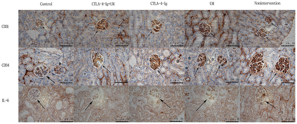
Figure 3.Analysis of renal CD31, CD34, and IL-6 staining. Analysis of renal CD31 staining. Control group: Weakly positive CD31 expression, with no evidence of blood vessel proliferation in the glomerulus (black arrow); CTLA-4-Ig + UM group: Strongly positive CD31 expression, with a small number of blood vessels in the glomerulus (black arrow); CTLA-4-Ig group: Strongly positive CD31 expression, with a small number of blood vessels in the glomerulus (black arrow); UM group: Strongly positive CD31 expression, with significant vascular proliferation in the glomerulus (black arrow); Non-intervention group: Strongly positive CD31 expression, with significant vascular proliferation in the glomerulus (black arrow). Analysis of renal CD34 staining. Control group: Weakly positive CD34 expression, with no evidence of blood vessel proliferation in the glomerulus (black arrow); CTLA-4-Ig + UM group: Strongly positive CD34 expression, with a small number of blood vessels in the glomerulus (black arrow); CTLA-4-Ig group: Strongly positive CD34 expression, with a small number of blood vessels in the glomerulus (black arrow); UM group: Strongly positive CD34 expression, with significant vascular proliferation in the glomerulus (black arrow); Non-intervention group: Strongly positive CD34 expression, with vascular hyperplasia in the glomerulus (black arrow). Analysis of renal IL-6 staining. Control group: Negative IL-6 expression in the glomerulus (black arrow); CTLA-4-Ig + UM group: Weakly positive IL-6 expression in the glomerulus (black arrow); CTLA-4-Ig group: Weakly positive IL-6 expression in the glomerulus (black arrow); UM group: Positive IL-6 expression in the glomerulus (black arrow); Non-intervention group: Strongly positive IL-6 expression in the glomerulus (black arrow).