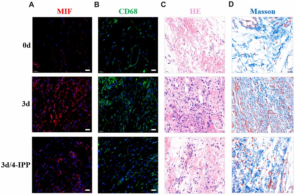
Figure 4.Inhibition of MIF in the lesion area attenuated posterior joint capsule inflammation and fibrosis. (A) Expression of MIF (red) in the posterior joint capsule was assessed via immunostaining at 0 d, 3 d and 3 d after injection of 4-IPP. (B) Immunostaining of CD68-positive macrophages (green) in the posterior joint capsule. (C) HE staining of the posterior joint capsule. (D) Masson staining of the posterior joint capsule. Scale bars, 20 μm.