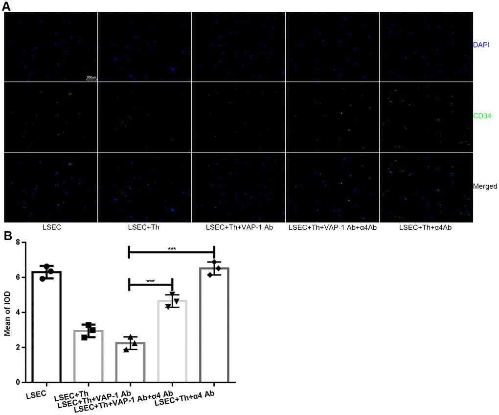
Figure 5.Expression of CD34 on LSECs in each group after in vitro coculture for 3 h. (A) Immunofluorescence staining for CD34 expression on LSECs (original magnification 100×). Anti-CD34 antibodies were labeled with Alexa Fluor 488 (green), and cell nuclei were labeled with DAPI (blue). (B) Quantification of CD34 expression of each group in (A). ***p < 0.001.