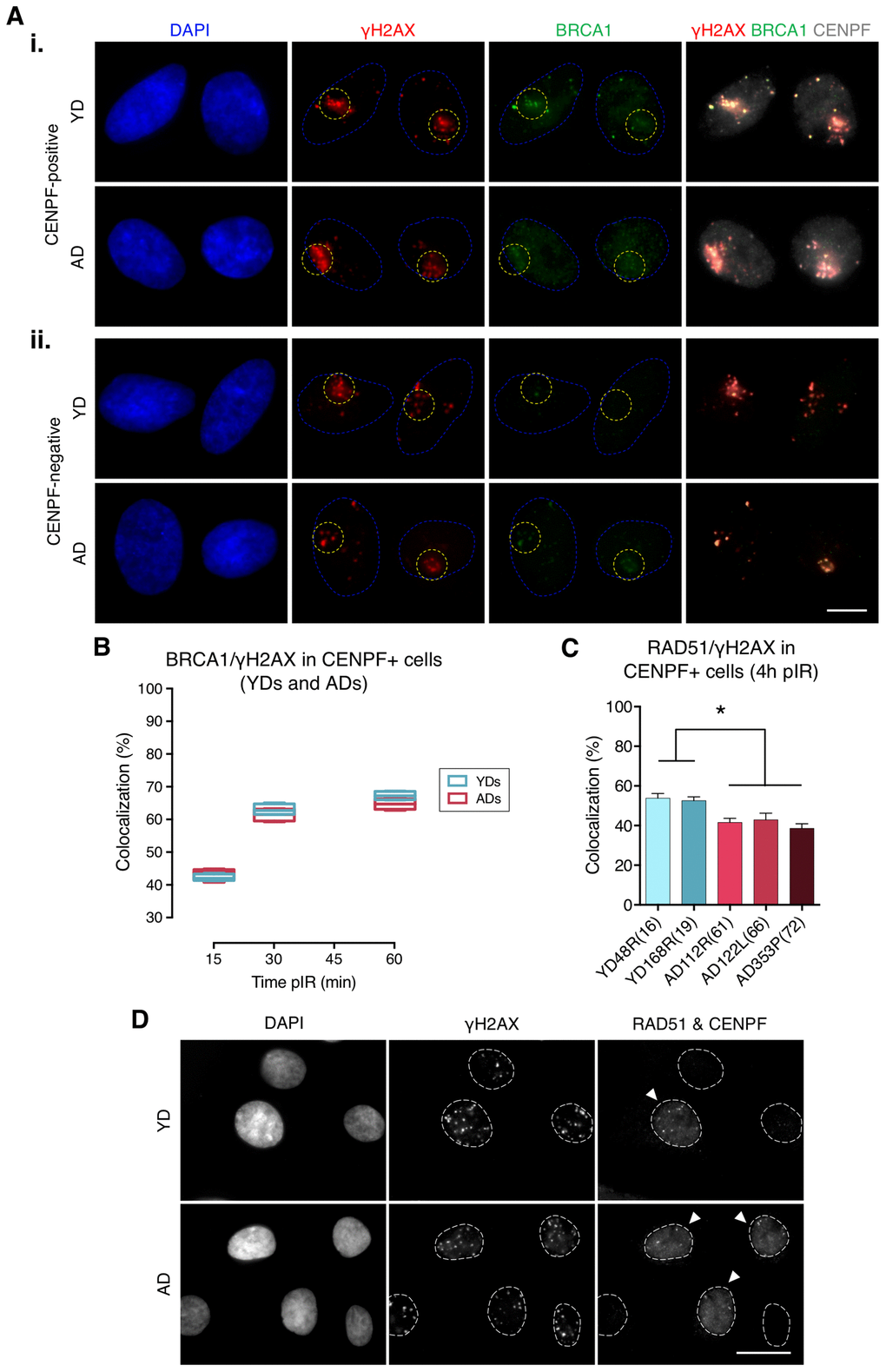
Figure 2.Recruitment of BRCA1 to DNA damage sites in G2 is not impaired in cells from aged donors. (A) Immunofluorescent labeling of cell nuclei (DAPI, blue), γH2AX (A594, red), BRCA1 (A488, green) and CENPF (A532, grey). γH2AX and BRCA1 foci were scored within the irradiated pore zone (yellow dotted lines) in CENPF-positive (i) or CENPF-negative (ii) cells. Scale bar = 10 μm. (B) Percentage of BRCA1/γH2AX foci colocalization in CENPF-positive HMECs from four YDs and four ADs. Boxes include data from the upper to the lower quartile and whiskers compile minimum to maximum values (n is stated in Supplementary Table 3). (C) Percentage of RAD51/γH2AX foci colocalization in CENPF-positive cells at 4 h after irradiation (5 Gy, γ-rays). Error bars indicate SEM (* p < .05; n ≥ 500 γH2AX foci/donor; one-way ANOVA + Tukey). (D) Immunofluorescent labeling of cell nuclei (DAPI), γH2AX (A488), RAD51 (A594) and CENPF nuclear staining (A532) at 4 h after exposure to 5 Gy of γ-rays. Arrowheads indicate G2 (CENPF-positive) cells. Scale bar = 20 μm.