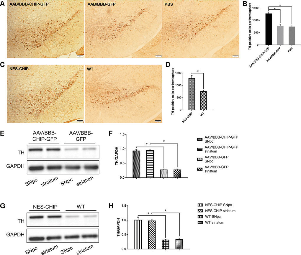
Figure 8.Immunohistochemistry of TH-positive cells and determine of TH protein. (A–D) Representative images of dopaminergic neurons stained for tyrosine hydroxylase (TH) in midbrain sections of different groups of mice. MPTP caused a significant decrease of TH-positive cells, while CHIP overexpression rescued the cell loss. (E–H) TH protein levels in SNpc and striatum of overexpression groups were significantly higher than those in control groups. *P<0.05.