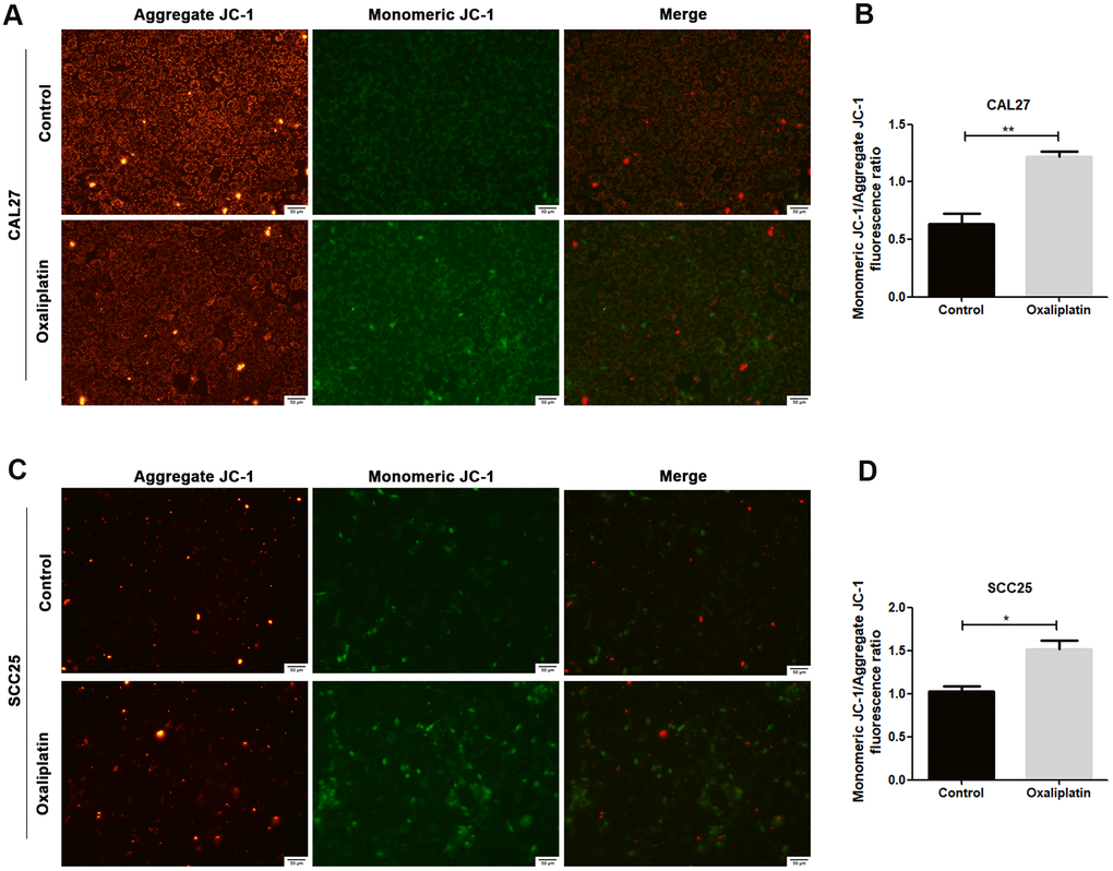
Figure 3.Oxaliplatin caused mitochondrial damage in OSCC cells. (A, C) Representative images of JC-1 staining in CAL27 and SCC25 cells treated with oxaliplatin. (B, D) The fluorescence ratio of monomeric JC-1/ aggregate JC-1 in CAL27 and SCC25 cells treated with oxaliplatin. * p<0.05, **p<0.01.