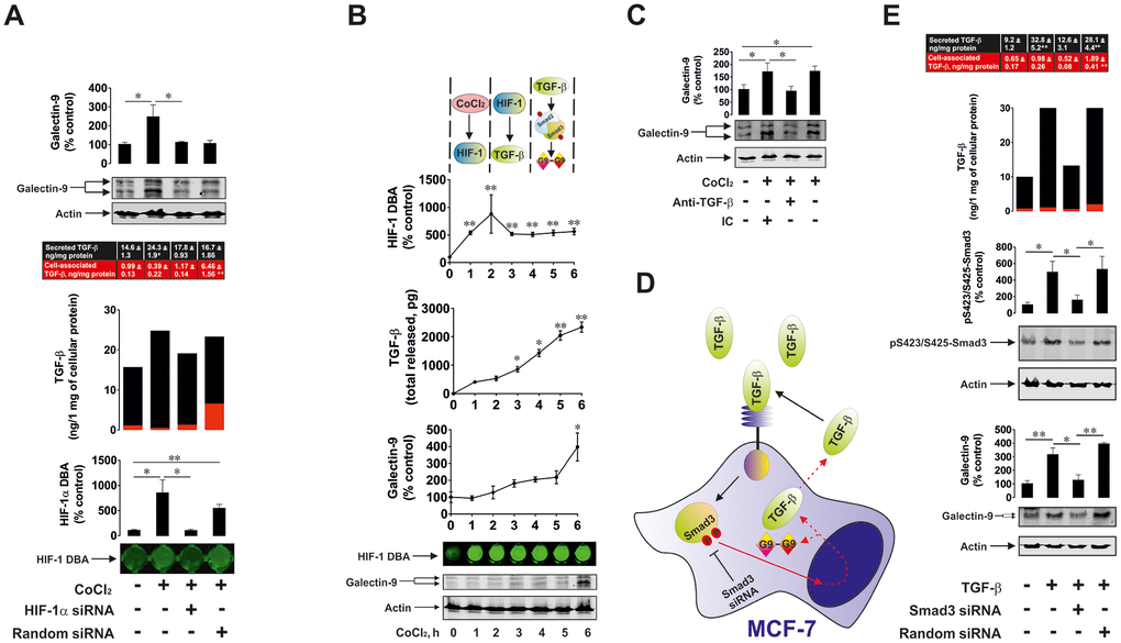
Figure 7.HIF-1 and Smad3 are involved in the production of TGF-β and galectin-9. (A) Cobalt chloride induces HIF-1 activation, TGF-β and galectin-9 production. Wild type, HIF-1α knockdown and random siRNA-transfected MCF-7 cells were exposed to 50 μM cobalt chloride for 6 h followed by measurement of HIF-1 DBA, secreted (in cell culture medium) and cell-associated (in cell lysates) TGF-β as well as cell-associated galectin-9. Images are from one experiment representative of three which gave similar results. (B) Dynamics of cobalt chloride-induced HIF-1 activation, TGF-β and galectin-9 accumulation in MCF-7 human breast cancer cells. MCF-7 cells were exposed to 50 μM cobalt chloride for 1, 2, 3, 4, 5 and 6 h followed by measurement of HIF-1 DBA, secreted and cell-associated TGF-β as well as cell-associated galectin-9. Images are from one experiment representative of three which gave similar results (in the case of TGF-β – vs 1 h time-point since at zero point cells can’t release any TGF-β. At this time-point, fresh medium has just been supplied and measurement was taken immediately to confirm zero TGF-β level). (C) HIF-1-induced galectin-9 expression is mediated by TGF-β. MCF-7 cells were exposed to 50 μM cobalt chloride for 6 h with or without presence of TGF-β neutralising or isotype control antibody. Galectin-9 expression was then assessed by Western blot. Images are from one experiment representative of three, which gave similar results. (D), (E) Smad3 is involved in TGF-β and galectin-9 expression. (D) Scheme of the experiment performed showing studied effects. (E) Wild type, Smad3 knockdown and random siRNA-transfected MCF-7 cells were exposed to 2 ng/ml TGF-β for 24 h followed by measurement of secreted (in cell culture medium) and cell-associated (in cell lysates) TGF-β as well as cell-associated galectin-9 and phospho-S423/S425 Smad3. Images are from one experiment representative of three, which gave similar results. All quantitative data are shown as mean values ± SEM (n=3-4) * - p < 0.05 and ** - p < 0.01 vs indicated events.