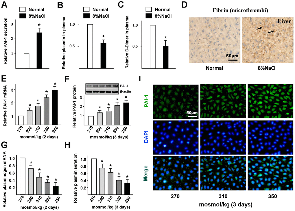
Figure 2.High-salt intake induces endothelial fibrinolytic dysfunction and thrombi. (A–C) Protein levels of PAI-1, active plasmin and D-Dimer in plasma of mice in normal and high salt groups after 4 weeks feeding. (D) Representative sections of the livers in ApoE-/- mice stained for fibrin. Nuclei, hematoxylin staining. Microthrombi were marked by arrowheads. (E, F) mRNA and protein expression of PAI-1 in HUVECs that cultured with different hyper-osmotic media (270, 290, 310, 330 and 350 mosmol/kg) for two or three days. 270 mosmol/kg was as the control. (G, H) mRNA and protein level of plasmin in HUVECs that cultured with different hyper-osmotic media for two or three days. 270 mosmol/kg was as the control. (I) Representative immunofluorescent staining of PAI-1 (green) in HUVECs that exposed to different hyper-osmotic media for three days. Nuclei were stained by DAPI. All data were presented as mean ± SEM, N≥3. *p < 0.05 versus control group.