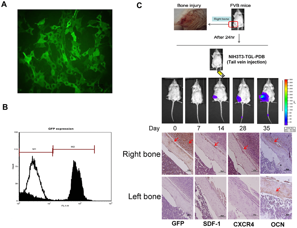
Figure 3.In vitro characterization and in vivo therapeutic effect of GFP transfected MEFs (NIH3T3) containing lentivirally transduced genes, thymidine kinase (T), green fluorescence protein (G) and luciferase genes (L) (NIH3T3-TGL). (A) GFP transfected NIH3T3 cell line were sorted out among other cells types by flow cytometry and the expression of GFP in MEF-TGL cells was validated by fluorescent microscopy exhibiting strong green signals (B). (C) Experimental presentation of femur bone injury created at mid-diaphysis of bilateral right femur, and cellular migration from administered site in FVB mice. Increased GFP-positive cells were detected at indicated time points. Histologic images also confirm the presence of GFP-positive signals (brown color) in right femur, while no signal appeared in left femur (control).