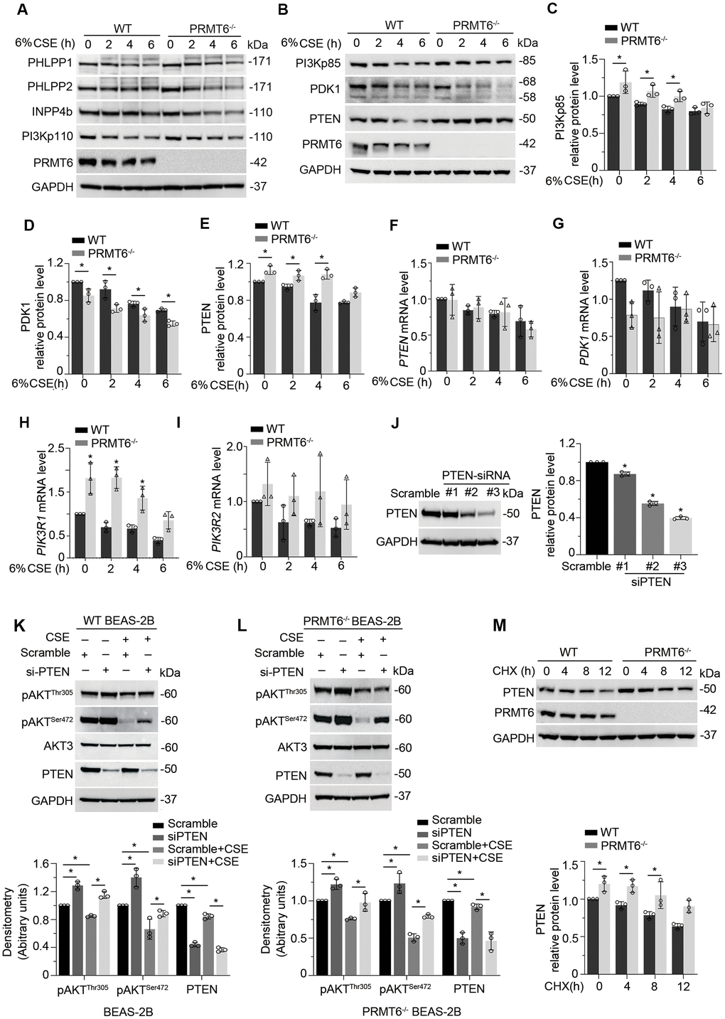
Figure 6.CSE suppresses AKT phosphorylation via PRMT6/PTEN signal transduction. (A, B) WT and PRMT6-/- BEAS-2B cells were treated with CSE at 2, 4, 6h. Western blotting was performed to detect the protein level of PHLPP1, PHLPP2, INPP4b, PI3Kp110, PI3Kp85, PDK1, PTEN, PRMT6 and GAPDH. (C–E) The plotted densitometry results of PI3Kp85, PDK1 and PTEN protein expression were presented. (F–I) WT and PRMT6-/- BEAS-2B cells were treated with CSE for 2, 4, 6h. qRT-PCR was performed to detect the mRNA level of PTEN, PDK1, PIK3R1 and PIK3R2. Results of qRT-PCR were shown as mean ± SD and representative of n=3 experiments. (J) Scramble siRNA and three kinds of double strands PTEN-siRNA were transfected into BEAS-2B separately. PTEN expression was detected by western blotting after transfection of PTEN-siRNA for 72hrs. Plotted PTEN protein level was presented in the right panel. (K, L) PTEN expression was silenced by DsiRNA in wild type and PRMT6 KO BEAS-2B cells. Western blotting was used to assay the AKT phosphorylation level. Representative blots of PTEN and PRMT6 were shown. (M) Cycloheximide (CHX, 100ug/ml) was applied to WT and PRMT6-/- BEAS-2B cells for 0, 4, 8, 12h. Densitometric results of PTEN and PRMT6 blots were plotted (lower panel). Results were representative of n=3 experiments. Statistics were measured by 1-way and 2-way ANOVA, *: p < 0.05.