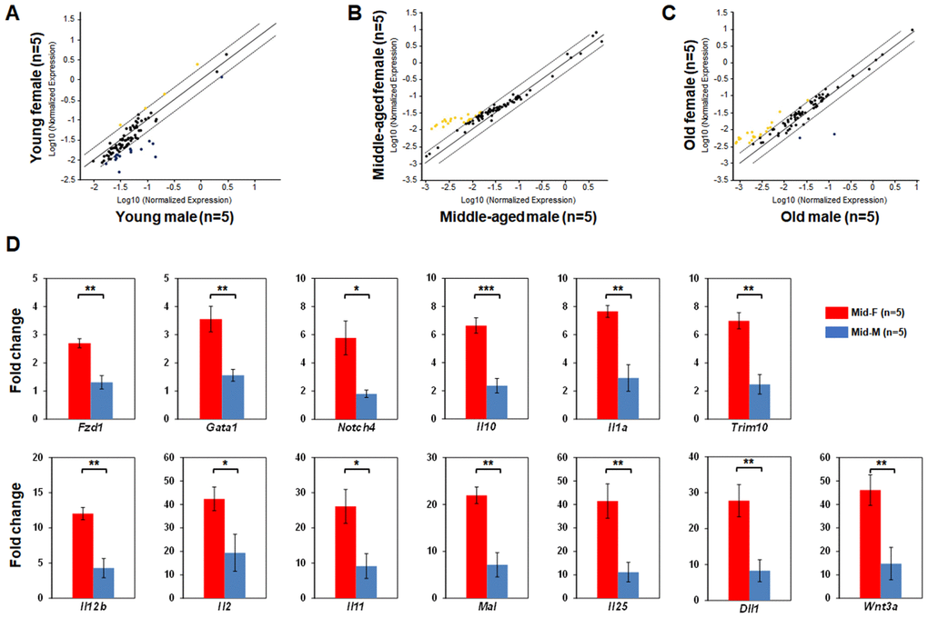
Figure 3.Comparisons of relative hematopoietic gene expression in BM-derived LSK cells between male and female of young, middle-aged and old mice. (A) Comparison between young female and young male mice. (B) Comparison between middle-aged female and middle-aged male mice. (C) Comparison between old female and old male mice. Yellow dots indicate genes with potentially higher level of expression in mice of y-axis than that in mice of x-axis; black dots indicate genes with comparable level of expression; blue dots indicate genes with potentially lower level of expression in mice of y-axis than that in mice of x-axis. (D) Real time PCR on individual genes represented by yellow dots from (B) was carried out to confirm that they had a significantly higher expression in LSK cells from middle-aged female than middle-aged male mice. * P < 0.05, ** P < 0.01, *** P < 0.001.