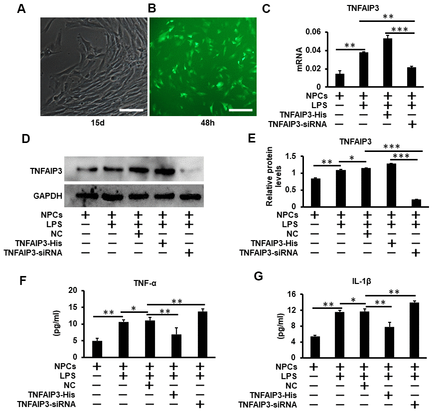
Figure 2.TNFAIP3 reduces pro-inflammatory cytokines expression in LPS-stimulated human NPCs. (A) Human primary nucleus pulposus cells isolated from patients with lumbar fractures (LVF), amplification × 100. (B) Human nucleus pulposus cells were transfected with Ad-TNFAIP3 for 48 h. (C–E) TNFAIP3 responses to inflammatory stimulation. After treatment with LPS for 24 h, the mRNA and protein expression levels of TNFAIP3 were measured by RT-qPCR and Western blot, respectively. (F, G) After transfected with TNFAIP3-His and TNFAIP3-siRNA for 24 h, then stimulated with LPS for 24 h, the expression levels of TNF-α, IL-1β were tested by ELISA. Data are represented by the mean ± standard deviation of 3 independent experiments. *** P<0.01, ** P<0.05, * P>0.05 by the Student t-test. Error bars indicate standard deviations. NC, negative control (adenovirus vector without TNFAIP3); TNFAIP3-His, Adenovirus with TNFAIP3 overexpression; TNFAIP3-siRNA, Adenovirus with TNFAIP3 knockdown. TNFAIP3-His +LPS, human NPCs transfected with TNFAIP3-His and then treated with LPS. TNFAIP3-siRNA +LPS, human NPCs transfected with TNFAIP3-siRNA and then treated with LPS.