Submit an Article
Navigate
Home
Editorial Board
Editorial Policies
Current Volume
Archive
Scientific Integrity
Publication Ethics Statements
Interviews with Outstanding Authors
Newsroom
Sponsored Conferences
Podcast
Contact
Special Collections
Submit an Article
Online ISSN: 1945-4589
Research Paper
|
Volume 12, Issue 23
|
pp. 24168–24183
Oxidative stress mediates age-related hypertrophy of ligamentum flavum by inducing inflammation, fibrosis, and apoptosis through activating Akt and MAPK pathways
Back to article
Figure 8
(8 of 8)
−
100%
+
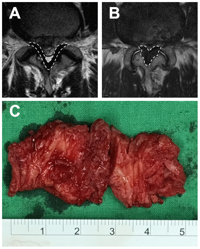
Figure 8.
MRI image and the LF specimen.
The T2-weighted image of the LF, surrounded by broken lines, at the L4/5 level of (
A
) an LDH patient and (
B
) an LSS patient. (
C
) A hypertrophic LF specimen.