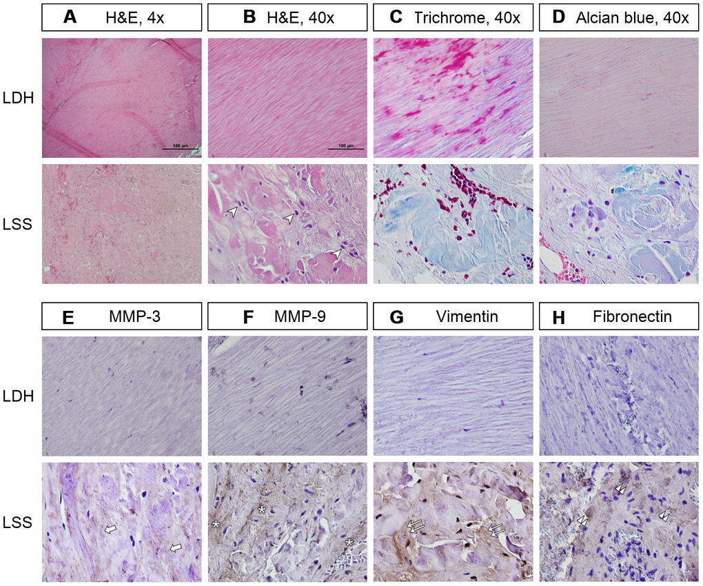
Figure 1.The LF of lumbar spinal stenosis (LSS) patients was positively stained for inflammatory cell infiltration and fibrotic markers. (A, B) In the LF of lumbar disc herniation (LDH) patients, the collagen fibers were well organized in a parallel order. In contrast, those from LSS patients showed signs of inflammation and fibrosis, including infiltration of inflammatory cells (arrowheads) and deposition of disorganized collagen fibers. Scale bar 100μm. (C) Masson’s trichrome staining revealed that the LF from LSS patients was stained a blue color with only traces of pink, further substantiating the increase in collagen deposition. (D) Alcian blue staining of the LF from LSS patients was stained a blue color, indicating increased deposition of glycosaminoglycans. (E, F) Immunohistochemical (IHC) analysis demonstrated that MMP-3 (arrows) and MMP-9 (asterisk) were positively stained on the LF specimens of LSS patients. (G, H) IHC analyses revealed that vimentin (double arrows) and fibronectin (double arrowheads) were positively stained on the LF specimens of LSS patients.