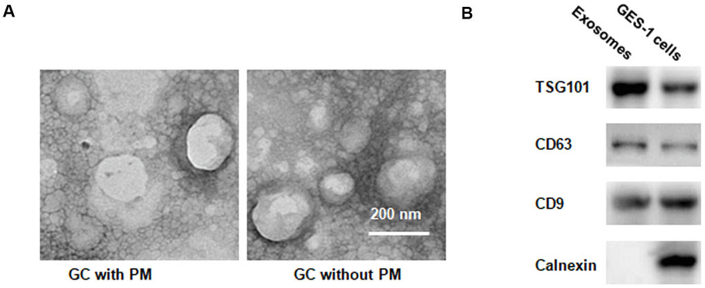
Figure 2.EVs identification in peritoneal fluid. (A) Transmission electron microscopy demonstrating many EVs <200 nm in diameter. (B) Western blotting illustrating the presence of TSG101 and CD63, two common EV markers, in EV fraction isolated from peritoneal fluid.