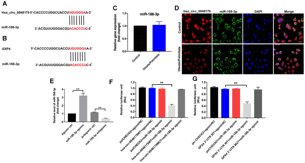
Figure 4.miR-188-3p forms a bridge connecting hsa_circ_0048179 and GPX4. (A) Sequence alignment of miR-188-3p with the putative binding sites within the WT regions of hsa_circ_0048179. (B) Sequence alignment of miR-188-3p with the putative binding sites within the WT regions of GPX4. (C) HepG2 cells were incubated with oleate/palmitate (2:1 molar ratio) for 24 h. after which levels of miR-188-3p expression were detected using qRT-PCR. (D) Cellular localization of hsa_circ_0048179 and miR-188-3p in HepG2 cells were analyzed using FISH assays. (E) HepG2 cells were transfected with miR-188-3p agomir or miR-188-3p antagomir for 72 h, after which levels of miR-188-3p expression were detected using qRT-PCR. **P<0.01 (F) Dual luciferase reporter assays were used to detect the luciferase activity in 293T cells following co-transfection with hsa_circ_0048179-WT/MUT 3’-UTR plasmid and miR-188-3p agomir. **P<0.01 (G) Dual luciferase reporter assays were used to detect luciferase activity in 293T cells after co-transfection of GPX4-WT/MUT 3′-UTR plasmid and miR-188-3p agomir. **P<0.01.