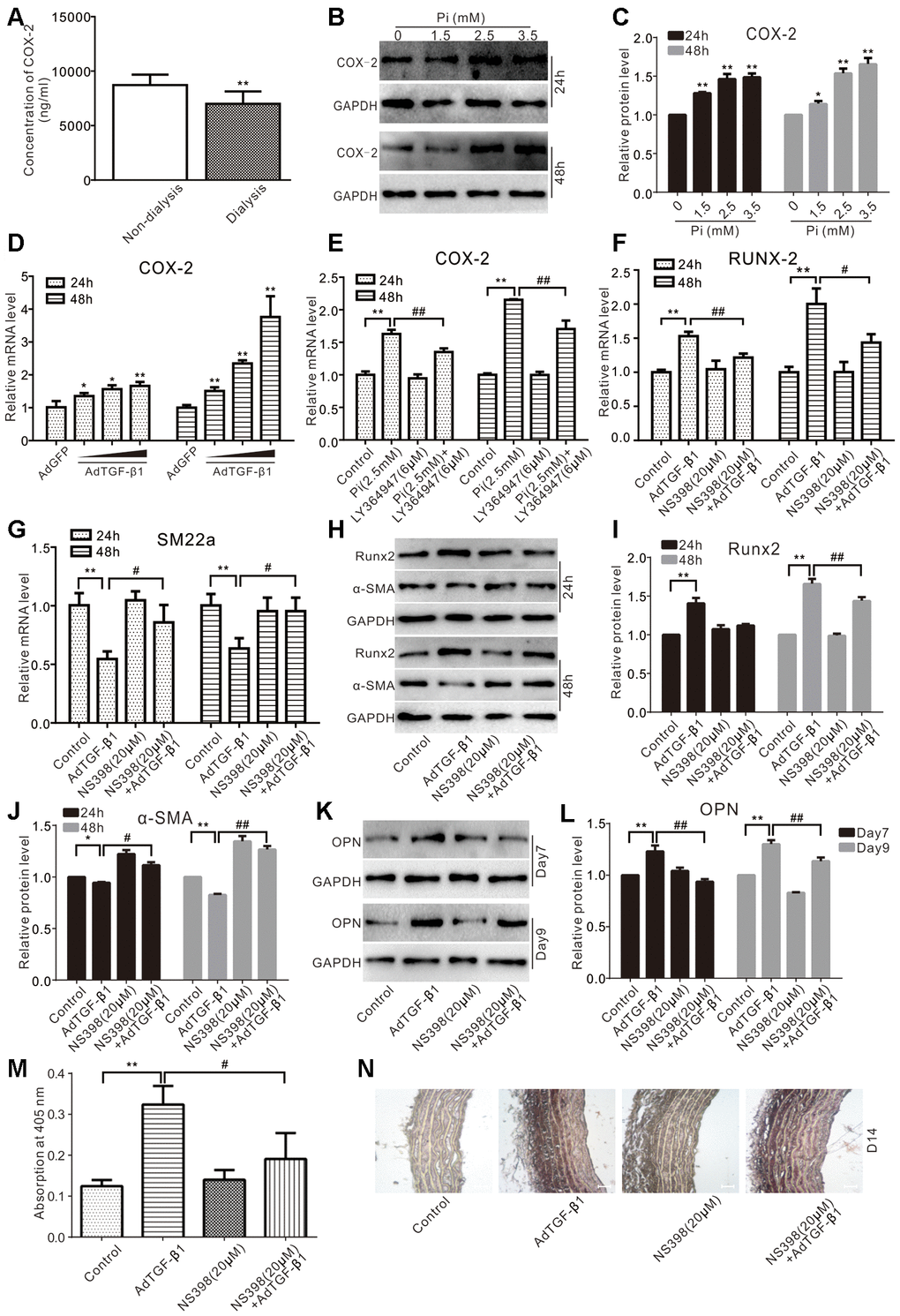
Figure 4.Effects of COX-2 on high phosphate and/or TGF-β1 induced calcification in VSMCs. (A) ELISA analysis results show the serum level of COX-2 in CKD patients treated with regular dialysis or non-dialysis (n=48) ("**" p<0.01 vs. non-dialysis). (B) Western blot assay results show the effect of high phosphate on COX-2 in VSMCs cells; GAPDH was used as a loading control. (C) Quantitative results of Western blot assay show the effect of high phosphate on COX-2 in VSMCs cells ("*" p<0.05 and "**" p<0.01 vs. control). (D) Real-time PCR assay results show the effect of TGF-β1 on mRNA expression of Cox-2 in VSMCs cells ("*" p<0.05 and "**" p<0.01 vs. control). (E) Real-time PCR assay results show the effect of high phosphate and/or LY364947 on mRNA expression of Cox-2 in VSMCs cells ("**" p<0.01 vs. control, "##" p<0.01 vs. high phosphate group). (F) Real-time PCR assay results show the effect of TGF-β1 and/or NS398 on expression mRNA of Runx-2 in VSMCs cells ("**" p<0.01 vs. control, "#" p<0.05 and "##” p<0.01 vs. TGF-β1 group). (G) Real-time PCR assay results show the effect of TGF-β1 and/or NS398 on mRNA expression of SM22α in VSMCs cells ("**" p<0.01 vs. control, "#" p<0.05 vs. TGF-β1 group). (H) Western blot assay results show the effect of TGF-β1 and/or NS398 on Runx2 and α-SMA in VSMCs cells, GAPDH was used as a loading control. (I) Quantitative results of Western blot assay show the effect of TGF-β1 and/or NS398 on Runx2 in VSMCs cells ("**" p<0.01 vs. control, "##" p<0.01 vs. TGF-β1 group). (J) Quantitative results of Western blot assay show the effect of TGF-β1 and/or NS398 on α-SMA in VSMCs cells ("*" p<0.05 and "**" p<0.01 vs. control, "#" p<0.05 and "##" p<0.01 vs. TGF-β1 group). (K) Western blot assay results show the effect of TGF-β1 and/or NS398 on OPN in VSMCs cells; GAPDH was used as a loading control. (L) Quantitative results of Western blot assay show the effect of TGF-β1 and/or NS398 on OPN in VSMCs cells ("**" p<0.01 vs. control, "##" p<0.01 vs. TGF-β1 group). (M) Quantitative analysis results of Alizarin Red S staining show the effect of TGF-β1 and/or NS398 on mineralization in VSMCs ("**" p<0.01 vs. control; "#" p<0.05 vs. TGF-β1 group). (N) The Von Kossa staining results show the effect of TGF-β1 and/or NS398 on calcification in aortic segments (Scale=100 μM). Pi: phosphate, LY364947: TGFβRI-specific inhibitor, NS398: COX-2-specific inhibitor.