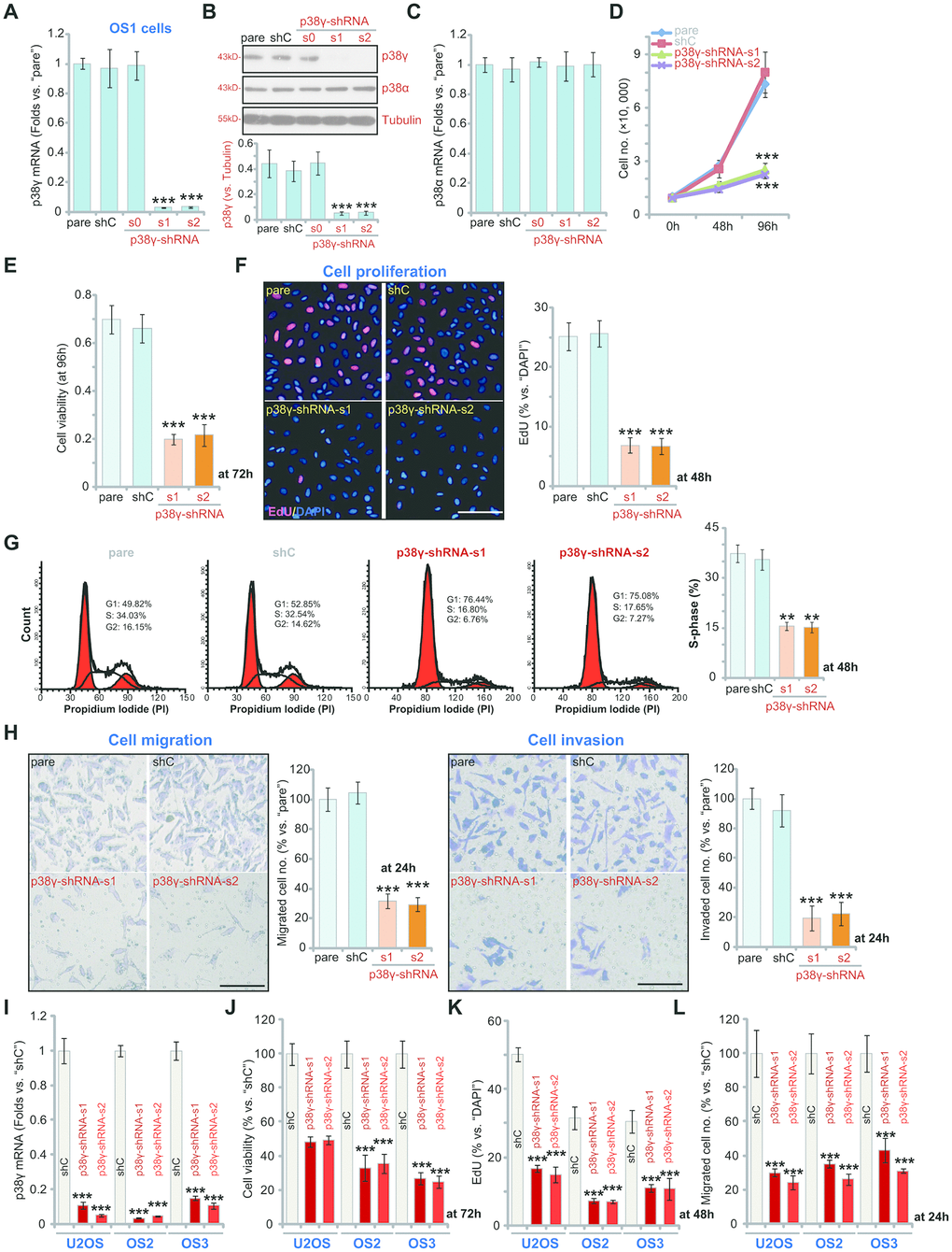
Figure 2.p38γ shRNA inhibits human OS cell viability, growth, proliferation, migration and invasion. Human OS cells, including OS1/OS2/OS3 primary OS cells (derived from three different OS patients) and the established U2OS cells, with scramble control shRNA (“shC”) or the applied p38γ shRNA (p38γ-shRNA-s0/s1/s2), were cultured and the expression of listed genes tested by qPCR and Western blotting assays (A–C, I); Cell growth (cell counting assay, D), viability (measuring CCK-8 viability OD, E, J) and proliferation (measuring EdU ratio, F, K) as well as cell cycle distribution (G), cell migration (“Transwell” assay, H, L) and invasion (“Matrigel Transwell” assay, H) were tested after incubation for applied time periods. “pare” indicated parental control cells (same for all Figures). For EdU staining assays, five random views with total 500 cell nuclei from each treatment were included to calculate the EdU/DAPI ratio (same for all Figures). For “Transwell”/“Martial Transwell” assays, in each condition five random views were included to calculate the average number of migrated/invaded cells (same for all Figures). For all the functional assays the same number of viable cells from the different genetic treatments were seeded initially onto each well or each dish (at 0h, same for all Figures). Expression of listed proteins was quantified and normalized to the loading control (B). Data presented as mean ± standard deviation (SD, n=5). ** p< 0.01 vs. “shC” cells. ** p< 0.001 vs. “shC” cells. Experiments in this figure were repeated five times. Bar=100 μm (F, H).