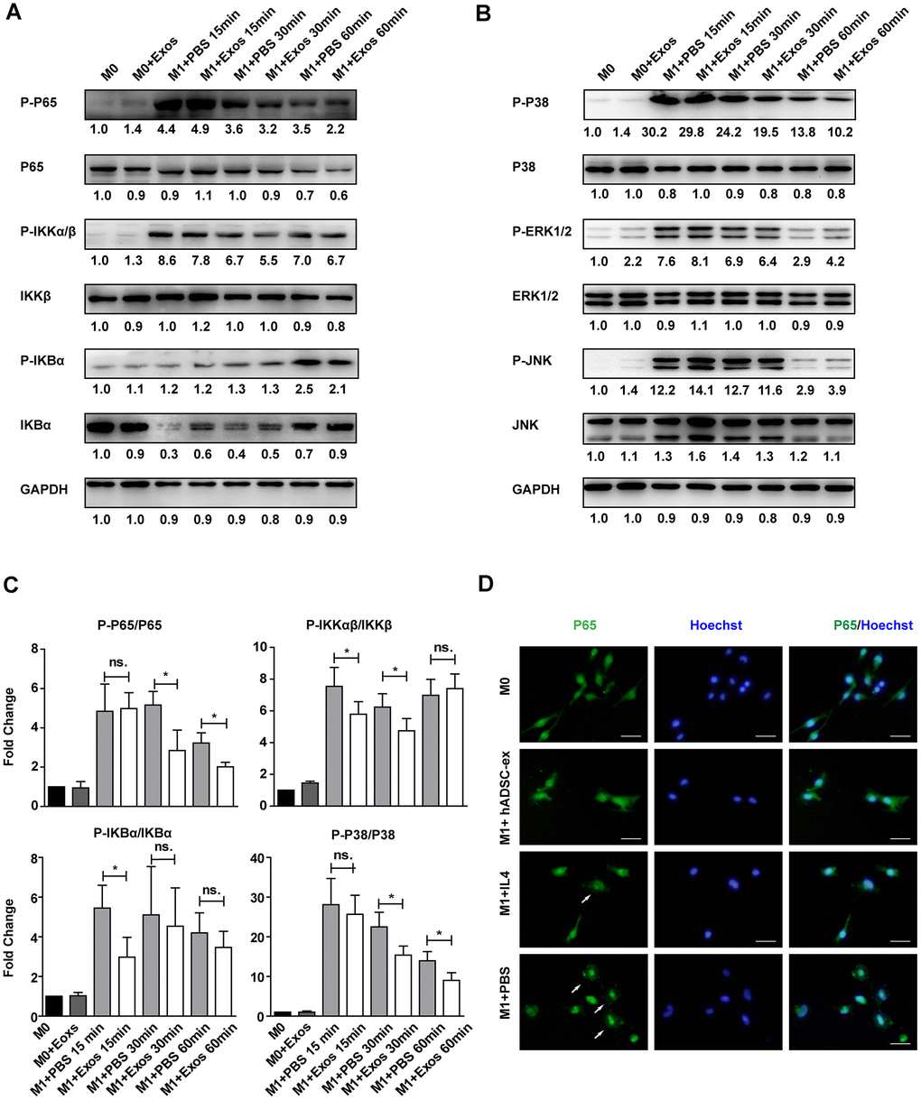
Figure 6.hADSC-ex suppressed the activation of classical NF-kB and MAPK signaling in primary microglia. (A, B) Immunoblots showing P-P65 and P65, P-IKKαβ and IKKβ, P-IKBα and IKBα, P-P38 and P38, P-ERK1/2 and ERK1/2, P-JNK and JNK, and GAPDH in M0 microglia cultured in M0 medium (containing 10 ng/mL M-CSF and 50 ng/mL transforming growth factor β1) or M1 medium, and treated with PBS or hADSC-ex (200 μg/mL) at different time points. (C) Fold changes in P-P65 to P65, P-IKKαβ to IKKβ, P-IKBα to IKBα, and P-P38 to P38 levels were each normalized to those of the M0 control group. Data represent the mean ± SD, n = 3 independent experiments, ns. p > 0.05, * p < 0.05, determined by one-way ANOVA vs. M1+PBS. (D) M0 microglia were cultured in M1 medium and treated with PBS, IL-4 (10 ng/mL) or hADSC-ex (200 μg/mL) for 24 h. The cells were stained for NF-κB P65 protein (green), and the nuclei were counterstained with Hoechst 33342 (blue). Scale bar = 50 μm. White arrows indicate the enrichment of nuclear NF-κB P65.