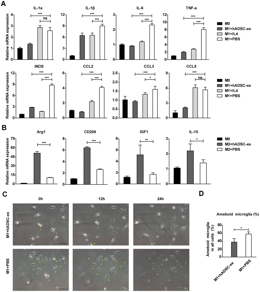
Figure 5.hADSC-ex alter the functional state of microglia in vitro. (A) qRT-PCR analysis of M1-associated factors after culture of M0 microglia in M1 medium (containing 10 ng/mL granulocyte M-CSF, 100 ng/mL lipopolysaccharide and 20 ng/mL interferon-γ) for 12 h with hADSC-ex (200 μg total protein/mL), IL-4 (10 ng/mL) or PBS. Data represent the mean ± SD, n = 3 independent experiments, ns. p > 0.05, * p < 0.05, ** p < 0.01, *** p < 0.001, determined by one-way ANOVA vs. M1+PBS. (B) qRT-PCR analysis of M2-associated factors after culture of M0 microglia in M2 culture medium (containing 10 ng/mL M-CSF and 10 ng/mL IL-4) for 12 h with hADSC-ex (200 μg total protein/mL) or PBS. Data represent the mean ± SD, n = 3 independent experiments, ns. p > 0.05, * p < 0.05, ** p < 0.01, *** p < 0.001, determined by one-way ANOVA vs. M2+PBS. (C) Representative images of morphological changes in microglia cultured in M1 medium for 24 h; yellow arrows indicate morphological changes. (D) The proportion of amoeba-like cells (M1 phenotype) among all cells after culture in M1 medium with hADSC-ex or PBS for 24 h. Data are presented as the mean ± SD, n = 3 independent experiments, * p < 0.05, determined by t-test vs. M1+PBS.