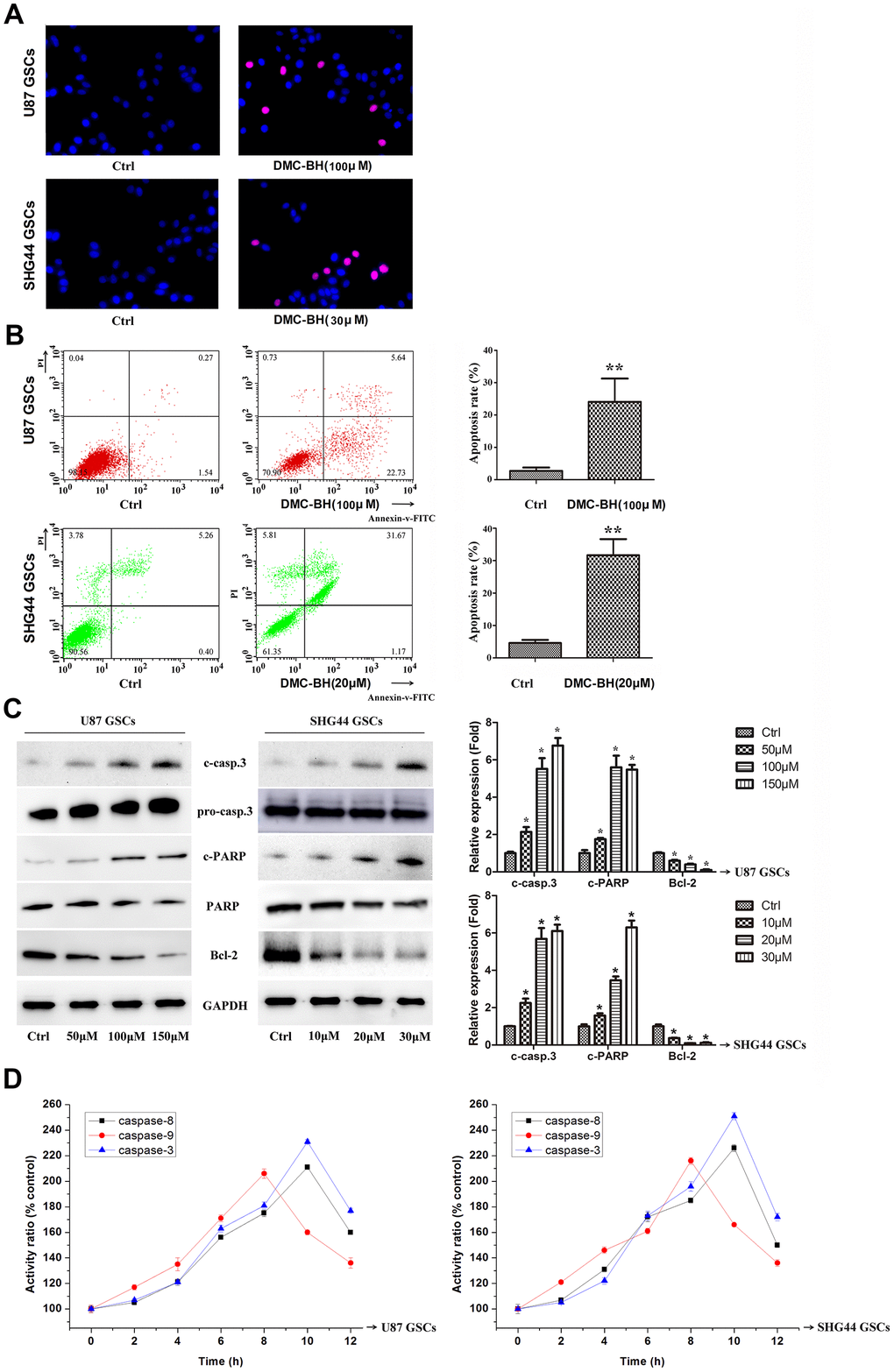
Figure 2.DMC-BH induces apoptosis in GSCs. (A) Morphological changes associated with apoptotic cell death analyzed by TUNEL staining. Red nuclear staining indicates apoptosis, while blue staining suggests normal nuclei. (B) Apoptosis analyzed using Annexin V-FITC/PI-staining flow cytometry. (C) Western analysis of apoptosis-related proteins caspase-3, PARP, and Bcl-2. (D) Caspase-3, -8 and -9 activity measured by ELISA.
(E) JC-1 positive cells analyzed by flow cytometry. (F) Intracellular ROS generation induced by 100 μM DMC-BH analyzed using DCFH-DA (10 μM) and flow cytometry. (G) Apoptosis of cells treated with DMC-BH and/or z-VAD-fmk for 24 h, analyzed by flow cytometry. (H) Caspase-3 activity measured using the substrate peptide Ac-DEVD-pNA; n =3.