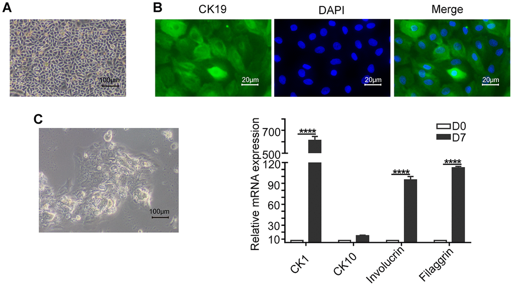
Figure 2.Basic characterization of keratinocytes. (A) Representative phase-contrast bright field image (scale bar: 100 μm) shows a confluent culture of the human skin keratinocytes. (B) Fluorescence images (scale bar: 20 μm) show positive expression of the epithelial stem cell marker, Cytokeratin 19 (CK19; green) in the keratinocytes. The nuclei are stained with DAPI (blue). (C) Representative phase-contrast bright field image (scale bar: 100 μm) shows agglomerate morphology of keratinocytes when grown in medium containing 1.2mM Ca2+ for 7 days. (D) Histogram plots shows the relative mRNA levels of differentiation markers CK1 (Cytokeratin 1), CK10 (Cytokeratin 10), Involucrin and Filaggrin levels in the keratinocytes on days 0 and 7. Note: The values are expressed as means ±SEM; ****p < 0.0001; ***p < 0.001; **p < 0.01; *p < 0.05.